Pyogenic granuloma is inflammatory granulation tissue and not a proliferation of tumor cells. It is a reactive lesion mainly composed of microvascular proliferation and inflammatory cell infiltration. The term pyogenic granuloma is not an appropriate one; pathologically, it is understood as an inflammatory reaction with excessive capillary proliferation, namely lobular capillary hemangioma.
This condition can occur at any age and is seen across a wide range from children to older adults. It most often appears as a complication of a chalazion and presents as a raised mass on the palpebral conjunctiva. It can also occur after eye surgery such as pterygium surgery, chalazion surgery, or strabismus surgery, and after eyelash removal or trauma.
Conjunctival pyogenic granuloma is relatively common among benign conjunctival lesions. It can occur in children as a secondary condition to chalazion, and it is not specific to any age group. Because it may enlarge rapidly, many patients and families seek care thinking it might be a malignant tumor, but the usual course is benign.
QIs pyogenic granuloma cancer?
A
Pyogenic granuloma is not a malignant tumor (cancer). It is not a tumorous lesion caused by proliferation of tumor cells, but a reactive capillary proliferation (lobular capillary hemangioma) in response to inflammatory stimulation. Because it looks red and grows rapidly, it may be suspected as malignant, but it is a benign lesion and there is no concern about metastasis or malignant transformation.
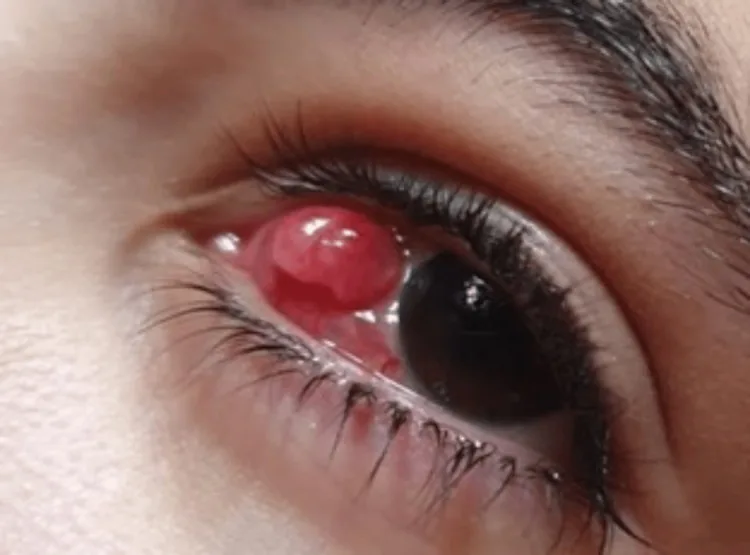
Bin Dlaim MS, Alhussein GA, Alqahtani RS, Almanea LT. Conservative Management of Giant Pyogenic Granuloma Post Strabismus Surgery: A Case Report and Literature Review. Cureus. 2023;15(7):e41321. Figure 1. PMCID: PMC10395757. License: CC BY.
A 5×8×5 mm pedunculated granuloma on the nasal conjunctiva that appeared 1 month after strabismus surgery; a bright red, shiny spherical mass protrudes from the conjunctival surface. This corresponds to the clinical appearance of the pedunculated red mass discussed in section “2. Main symptoms and clinical findings”.
A red, pedunculated, smooth, spherical to papillomatous mass appears on the palpebral conjunctival surface. The characteristic features of the mass are as follows.
Color: Bright red to dark red. Fine blood vessels run over the surface
Shape: Pedunculated (projecting from the conjunctiva on a stalk). Spherical to papillomatous
Surface: Smooth and glossy. Ulceration is usually not seen
Growth rate: It may enlarge rapidly. In some cases, it can reach several mm to over 1 cm within a few weeks
Bleeds easily: Because the mass contains abundant capillaries, it bleeds with slight contact
In cases with chalazion, the course often follows a pattern in which the mass protrudes from the conjunctival surface after the chalazion is incised or ruptures spontaneously. The finding of a pyogenic granuloma bulging outward from the chalazion contents is characteristic of this disease.
QWhat does a pyogenic granuloma look like?
A
It appears as a bright red, pedunculated mass on the conjunctival surface when the eyelid is turned inside out (palpebral conjunctiva). The surface is smooth and glossy, and it bleeds easily when touched. It may grow quickly and can reach nearly 1 cm within a few weeks. If it is associated with a chalazion, the mass may be seen protruding from the chalazion contents.
Chalazion (most common): Most cases occur after incision and drainage of a chalazion or after it ruptures spontaneously. A foreign-body reaction to meibomian gland lipid is thought to trigger capillary proliferation
After eye surgery: It is seen relatively often after pterygium surgery, chalazion surgery, and esotropia surgery (operations involving a conjunctival incision). After pterygium surgery, it is known to arise from the conjunctival suture site at the excision area
After trauma: It can develop after eyelash removal, after a foreign body enters, or after other conjunctival injuries
After infection: It may occur secondary to conjunctivitis or keratoconjunctivitis
Idiopathic: In some cases, no clear trigger can be identified
Pyogenic granuloma can often be diagnosed based on clinical findings alone. In typical cases, the course and appearance of a ‘rapidly enlarging pedunculated red mass after chalazion surgery’ are enough to make a clinical diagnosis.
Visual inspection and slit-lamp examination: The eyelid is everted, and the mass is assessed for color, shape, whether it is pedunculated, and surface characteristics
Check for association with chalazion: During the history, confirm the presence of chalazion and any history of incision or surgery
Pathology of the excised specimen: A pathological examination of the removed tissue is needed for a definitive diagnosis. If malignancy cannot be ruled out clinically, always send it for pathology
Common in children. Bluish raised mass. Tends to regress naturally after 1 year of age
Kaposi sarcoma
Seen in patients with immunodeficiency (HIV infection). Multiple purplish-red masses
Conjunctival metastatic tumor
Should be ruled out if there is a history of malignant tumor
If the differential diagnosis is difficult, or if atypical findings such as rapid growth, recurrence, or a firm texture are present, actively excise the lesion and confirm the diagnosis with pathological examination.
Treatment for pyogenic granuloma is chosen based on the size of the lesion, the trigger, and the patient’s condition. The main treatment options are shown below.
Observation
Indications: Relatively small lesions. When waiting for spontaneous sloughing after treating an inflammatory trigger (such as a chalazion).
Points to note: If the lesion grows rapidly or bleeds easily, continued observation may be difficult.
Course: It may fall off on its own. It may shrink or disappear after the chalazion is treated at the same time.
Local steroid therapy
Eye drops: Betamethasone eye drops (0.1%) or fluorometholone eye drops (0.1%) are given 4 to 6 times a day.
Subconjunctival injection: Triamcinolone acetonide (4 mg/mL) may be injected subconjunctivally at 0.2 to 0.5 mL. A reduction in the mass can be expected.
Points to note: Use while checking intraocular pressure regularly. Be careful about steroid-induced glaucoma.
Excision
Indications: Persistent lesions, large lesions, cases with an insufficient response to steroids, or when malignancy cannot be ruled out.
Procedure: Under local anesthesia, the lesion is excised from the base of the stalk. The excised specimen is submitted for pathological examination.
When chalazion is present: Perform incision and drainage of the chalazion at the same time. If the chalazion remains, the risk of recurrence increases.
To prevent recurrence after excision, complete removal of the causative chalazion is important. If the chalazion remains, pyogenic granuloma can recur in the same area even after it is removed. In recurrent cases, repeat excision is performed, and the excised specimen is sent for pathological examination to rule out malignant disease.
QCan treating the chalazion cure the pyogenic granuloma?
A
In pyogenic granuloma that develops secondary to a chalazion, incision and drainage treatment of the chalazion may reduce or eliminate the pyogenic granuloma. However, if the pyogenic granuloma has become large, treatment of the chalazion alone often does not improve it, and excision of the pyogenic granuloma itself is needed. Treating the chalazion and pyogenic granuloma at the same time makes recurrence less likely.
QCan it recur after treatment?
A
Recurrence after excision can occur. The risk is especially high if a chalazion remains at the excision site. If recurrence occurs, repeat excision is performed, and the excised specimen is sent for pathological examination to confirm the differential diagnosis from malignant disease. Even when treated with steroids alone, if the triggering chalazion remains, the condition can flare up again.
6. Pathophysiology and detailed mechanism of onset
Pyogenic granuloma develops because of an excessive vascular proliferative response of conjunctival tissue to inflammatory stimuli such as trauma, surgery, and chalazion. Pathologically, it is not a tumor but an inflammatory lesion accompanied by angiogenesis.
Persistent local inflammation leads to production of angiogenic factors, including vascular endothelial growth factor (VEGF). These factors promote the formation and proliferation of new capillaries, resulting in a lobular capillary hemangioma. Lobular proliferation of capillaries is seen within edematous stroma accompanied by infiltration of inflammatory cells (neutrophils, lymphocytes, and plasma cells).
In chalazion, a foreign body granuloma centered on macrophages forms in response to lipid components leaking from an obstructed meibomian gland. This foreign body reaction can protrude onto the conjunctival surface and trigger pyogenic granuloma. If the chalazion contents are not completely removed after incision and drainage, the foreign body stimulus persists and the pyogenic granuloma may recur.
When pyogenic granuloma develops after surgery involving a conjunctival incision, such as pterygium surgery or strabismus surgery, the presumed mechanisms include a foreign body reaction to the suture material and excessive vascular proliferation at the incision site. Cases arising around sutures, especially absorbable sutures, have been reported 1), and the lesion may shrink or disappear after suture removal.
7. Latest research and future prospects (research-stage reports)
Comparative studies have been conducted on the effectiveness of steroid eye drops and subconjunctival injection (triamcinolone acetonide). Subconjunctival injection can achieve a higher local concentration than eye drops, so a more reliable shrinkage effect is expected, but it carries risks of pain, increased intraocular pressure, and depigmentation, making patient selection important 2).
Pediatric pyogenic granuloma is often associated with chalazion, and anti-inflammatory eye drops (such as fluorometholone) are tried as initial treatment; excision is selected for cases with poor response. General anesthesia may be required, and the treatment plan is determined by considering age, lesion size, and the child’s level of cooperation 3).
Systematic data on the spontaneous regression rate and time to regression of pyogenic granuloma are limited. It is known that small lesions may fall off spontaneously within a few weeks to a few months, but further research is needed on the characteristics and predictive factors of lesions for which spontaneous regression can be expected4).
Shields JA, Shields CL, Eagle RC Jr, et al. Pyogenic granuloma of conjunctiva. Arch Ophthalmol. 1995;113(12):1555-1558.
Ferry AP.. Pyogenic granulomas of the eye and ocular adnexa: a study of 100 cases. Trans Am Ophthalmol Soc. 1989;87:327-43; discussion 343-7. PMID:2562522; PMCID:PMC1298549.
Rios JD, Dohlman CH, Tomlinson A, et al. Conjunctival pyogenic granuloma in children. J Pediatr Ophthalmol Strabismus. 2002;39(5):293-296.
Reddy SC, Reddy RC. Pyogenic granuloma of conjunctiva. Int J Ophthalmol. 2012;5(5):651-653.
Copy the article text and paste it into your preferred AI assistant.
Article copied to clipboard
Open an AI assistant below and paste the copied text into the chat box.