Enucleation is a surgical procedure that removes the entire eyeball and its intraocular contents while preserving the structures around the orbit (extraocular muscles, eyelids, and orbital fat).
Because there are procedures similar to those involving the eyeball, each definition needs to be clearly stated.
Enucleation (enucleation): Removes the entire eyeball and part of the optic nerve. The surrounding orbital structures are preserved.
Evisceration (evisceration): Preserves the sclera with the extraocular muscles attached and removes only the intraocular tissues. It has a slight cosmetic advantage, but is contraindicated when a malignant tumor is suspected.
Exenteration (exenteration): Removes all orbital contents, including the eyeball and soft tissue. It is a more extensive procedure.
It was first reported in the 1500s as “extirpation”; at that time, the conjunctiva and extraocular muscles were not preserved. In the mid-1800s, enucleation without an implant was described in the literature, and the first reports of implant insertion appeared in 1886-1887.
Histopathological examination of the removed eye is possible, and in intraocular malignant tumors the presence or absence of extraocular extension can be confirmed pathologically. Unlike evisceration, the entire eye is removed, so there is no risk of tumor seeding.
QHow is enucleation different from evisceration?
A
Enucleation removes the entire eye, whereas evisceration preserves the sclera and extraocular muscles and removes only the contents of the eye. Evisceration gives slightly better cosmetic results and better prosthesis mobility, but it is contraindicated when a malignant tumor is suspected, in which case enucleation is chosen. Enucleation also allows histopathological examination of the removed eye.
Uveal melanoma: The most common primary malignant intraocular tumor in adults. Indicated when eye-preserving treatment is unlikely to succeed. Assessment for optic nerve invasion is essential.
Retinoblastoma: A pediatric retinal malignant tumor caused by RB1 gene abnormalities. Enucleation is indicated when optic nerve invasion is suspected.
Traumatic and painful eye
Irreparable ocular trauma: Severe scleral damage with marked uveal prolapse, or a long delay between injury and medical care.
Painful phthisis bulbi: Indicated only when an intraocular tumor has been ruled out.
Absolute glaucoma: End-stage glaucoma that is resistant to medication and surgery.
Other
Prevention of sympathetic ophthalmia: When severe globe laceration or rupture is judged unreparable.
Severe infection: Advanced endophthalmitis with no prospect of visual recovery.
Microphthalmos: May be indicated for fitting an ocular prosthesis.
Considerations for primary enucleation in acute trauma
The classic “14-day rule” (removal within 14 days after trauma) has been shown to be arbitrary and to lack scientific support.
QIs an injured eye always going to need to be removed?
A
Not necessarily. Many surgeons first perform primary closure and consider enucleation if light perception is still absent afterward. After acute trauma, it is important to give the patient time to weigh the pros and cons. It has also been shown that the classic “14-day rule,” which says enucleation within 14 days can prevent sympathetic ophthalmia, has no scientific basis.
The main underlying diseases and risk factors that may require enucleation are shown below.
Intraocular malignant tumors: Choroidal melanoma is the most common primary intraocular malignant tumor in adults, and the main treatments are radiotherapy and enucleation. Retinoblastoma is a malignant retinal tumor in children caused by RB1 gene abnormalities, and enucleation is indicated when optic nerve invasion is suspected.
B-mode ultrasonography: Used to check for findings characteristic of choroidal melanoma (choroidal excavation, mushroom shape) and to assess for calcification seen in retinoblastoma and painful phthisis bulbi.
Contrast-enhanced MRI (contrast-enhanced CT if MRI is not suitable): Assesses whether a tumor is present, its size, and whether there is extraocular extension. It is essential for confirming an intraocular malignancy.
Intraoperative rapid pathology diagnosis: In enucleation for malignant tumors, rapid pathology is used to confirm whether tumor cells have infiltrated the cut end of the optic nerve of the removed eye. Confirming a negative margin means the treatment is complete.
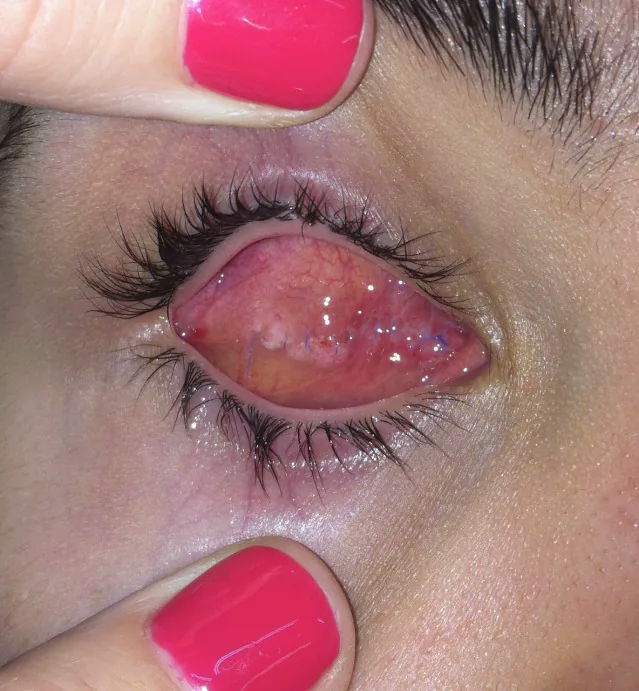
Schmitzer S, Simionescu C, Alexandrescu C, Burcea M. The Anophthalmic Socket - Reconstruction Options. J Med Life. 2014;7(Spec Iss 4):23-29. Figure 3a. PMCID: PMC4962761. License: CC BY.
This is a postoperative photograph of the orbit with a methyl methacrylate sphere placed within the muscle cone. It shows the coverage of the conjunctival surface and the positional relationship of the implant supporting the prosthetic eye bed.
It is often performed under general anesthesia. The conjunctiva is circumferentially incised at the limbus, and the sclera is exposed up to around the equator.
For the four rectus muscles, place a 5-0 PGA absorbable suture, cut the tendons, and separate them from the sclera. The key is to cut the medial and lateral rectus muscles while leaving enough tendon to hold the globe. Cut the superior oblique tendon and the inferior oblique muscle, and achieve thorough hemostasis at the cut ends.
Subluxation of the globe and optic nerve transection
After separating the posterior Tenon’s capsule from the sclera, grasp the insertion sites of the medial and lateral rectus muscles and gently rotate the globe while lifting it to subluxate it. This makes access to the optic nerve easier.
Insert the enucleation scissors along the sclera toward the back with the blades closed. Once the tip of the scissors feels the optic nerve as a cord-like structure, open the scissors slightly and cut the optic nerve in one motion as far back as possible. In cases of malignant tumors, try to cut a longer segment of the optic nerve.
Apply pressure with a finger immediately after cutting. Bleeding usually stops within 3 to 5 minutes. In malignant tumors, perform rapid pathological examination to check whether tumor cells have infiltrated the cut end of the optic nerve.
Insert a spherical implant (orbital implant) within the muscle cone, and suture the superior and inferior rectus muscles and the medial and lateral rectus muscles over the front of the implant.
Suture the anterior Tenon’s capsule and conjunctiva separately. Place antibiotic ointment in the conjunctival sac, insert a conformer, and apply a light pressure dressing.
Histopathology is possible: The entire removed eye can be examined histologically. It is the only option for malignant tumors.
Lower risk of sympathetic ophthalmia: Complete removal of exposure to uveal antigens (classic view).
More likely to cause a sunken eye: In a U.S. survey, 94% of ocularists said that a sunken eye and a deep upper eyelid sulcus are more common after enucleation.
Features of evisceration
Slight cosmetic advantage: Because the sclera and extraocular muscles are preserved, prosthesis movement is better.
Less sunken eye: In a survey of U.S.-certified ocularists, 82% said that evisceration gives the best cosmetic results.
Contraindicated: Should not be performed when malignancy is suspected.
As a criterion for choosing the procedure, if an early visit means the uveal tissue can be sufficiently removed under an operating microscope, evisceration is selected. If there is severe scleral damage or uveal prolapse, or if a long time has passed between injury and the visit, enucleation is selected.
The types and features of implants are shown below.
Type
Representative material
Features
Porous implant
Hydroxyapatite and porous polyethylene
Promotes fibrovascular tissue ingrowth. Extraocular muscle fixation is possible
Non-porous implant
Glass, silicone, acrylic, PMMA
Advantageous in situations with a high risk of infection. In Japan, PMMA resin spheres are the standard example
Porous polyethylene: The surface is smooth and no wrapping is needed. Extraocular muscles can be sutured directly, and it is cheaper than hydroxyapatite.
Hydroxyapatite: Introduced in 1989. Because the surface is rough, wrapping with donor sclera, acellular dermis, pericardium, and similar materials is needed.
Current situation in Japan: There is no orbital implant approved by the Ministry of Health, Labour and Welfare in Japan. PMMA resin spheres are the most common, and early approval and introduction are desired.
Implant size: A minimum of 20 mm is recommended. The formula “axial length minus 2 mm” is considered useful for supplementing orbital volume. The motility of the ocular prosthesis is affected more by implant size than by implant material, and larger implants provide better motility.
Short-term postoperative care: Wear an eye shield for a short period after surgery and return for follow-up one week later. Pain medicine and anti-nausea medicine are prescribed. There is not enough evidence to support preventive antibiotics, but some surgeons still prescribe them.
Wearing a conformer: If left for a long time, the conjunctival sac can shrink, so a conformer or ready-made prosthetic eye should always be worn.
Starting prosthetic eye wear: Begin wearing a prosthetic eye 1 to 2 months after surgery, once the conjunctival sac has settled. As a guide, prosthetic eye fabrication may begin 2 to 4 weeks after surgery, when pain and inflammation have eased.
Children: After congenital anophthalmia or removal in infancy, a prosthetic eye should be worn as early as possible to promote eyelid and orbital development (especially important under age 5).
QWhen can a prosthetic eye be worn?
A
Prosthetic eye wear is started 1 to 2 months after surgery, once the conjunctival sac has stabilized. Until then, a conformer (temporary prosthetic eye) must be worn to prevent the conjunctival sac from shrinking. If left for a long time, the conjunctival sac can shrink markedly, so early use of a conformer is important. In children, even earlier use is recommended for orbital growth.
Autologous dermis-fat grafting may be used for primary or secondary orbital reconstruction. In children, especially those under 5 years old, it is advantageous for promoting orbital growth and is suitable for restoring orbital volume. In children after enucleation for retinoblastoma, debulking may be needed during follow-up. For implants larger than 22 mm, be aware of the risk of early volume loss due to initial tension.
Basis for pathological diagnosis: By removing the entire eyeball and optic nerve, the depth of tumor invasion, whether tumor cells remain at the optic nerve margin, and whether there is extrascleral extension can be assessed histologically. This is not possible with evisceration.
Porous implants: The porous structure promotes fibrovascular ingrowth, and tissue integration improves motility. Fixation to the extraocular muscles is also better.
Non-porous implants: Because there is no tissue ingrowth, motility may be reduced and the risk of implant migration may be higher.
After enucleation, loss of orbital volume can cause superior sulcus deepening, enophthalmos, and ptosis. This condition is called “post-enucleation socket syndrome.” Inadequate fixation of the extraocular muscles to the implant can cause implant migration and worsen symptoms. Choosing an implant of appropriate size and securely suturing the extraocular muscles are key to prevention.
7. Latest research and future prospects (research-stage reports)
Visual hallucinations called Charles Bonnet syndrome (CBS) can occur after enucleation. CBS was traditionally thought to require a loss of more than 60% of binocular vision to develop, but it has been shown that it can also occur with loss of vision in one eye only.
Forte et al. (2025) reported a 67-year-old woman who developed CBS after enucleation for choroidal melanoma1). Visual hallucinations began the day after surgery and continued for 2 years. Vision in the remaining eye was -0.07 LogMAR (good), and mean sensitivity on microperimetry was 24.7 dB. The hallucinations included moving patterns, colored flashes, fire, Christmas trees, robots, and kingfishers, occurring several times a week and lasting at least 10 seconds each time. The patient recognized that the hallucinations were not real (insight preserved).
A literature review identified 9 cases of CBS after loss of vision in one eye. The mean age at diagnosis was 69.4 years (range 52-82), male-to-female ratio 3:1. In 8 of 9 cases, hallucinations began within hours to 2 days after vision loss, and in 7 of 9 cases they resolved (mean 17.6 days, range 2 hours-96 days). Complex hallucinations were seen in 100% of cases, and people were the most common images (56%)1).
It is recommended to explain the possibility of CBS to all patients undergoing enucleation before surgery and to screen for it after surgery.
QCan hallucinations occur after enucleation?
A
Yes. Visual hallucinations called Charles Bonnet syndrome (CBS) can occur. The hallucinations may include moving patterns, colors, and people, and the patient recognizes that they are not real (insight is preserved). It has been reported that CBS can occur even with loss of vision in one eye only1), so it is important to explain this to patients before surgery.
Traumatic enucleation and intracranial complications
In complete globe avulsion associated with self-inflicted eye injury (Oedipism) in patients with mental illness, subarachnoid hemorrhage (SAH) can occur because of optic nerve transection. Because the ophthalmic artery branches from the C6 segment of the internal carotid artery and runs through the subarachnoid space, rupture of the ophthalmic artery may cause SAH and internal carotid artery dissection.
Flippin et al. (2023) reported a case of bilateral self-enucleation in a patient with mental illness 2). Both globes were completely avulsed with part of the optic nerve attached. Head CTA showed suprasellar SAH with intraventricular extension and intraventricular hemorrhage (IVH). There were no facial fractures or vascular dissections, and after management in the trauma ICU, the ophthalmology team performed irrigation, debridement, orbital implant insertion, and closure. After 4 days in hospital, there were no neurological or ophthalmic complications, and the patient was admitted to psychiatry after discharge.
In cases with complete globe avulsion and optic nerve transection, head CT (preferably CTA) must be used to assess for intracranial hemorrhage.
It has been reported that daytime sleepiness from obstructive sleep apnea (OSA) can cause severe eye trauma and globe loss.
Baker et al. (2024) reported a case of a 55-year-old woman with OSA and obesity hypoventilation syndrome who suddenly fell asleep in the bathroom and struck a sharp object, resulting in right globe avulsion (about 5 cm optic nerve rupture) and enucleation with orbital implant insertion 3). The AHI was 55.6 events/hour and the lowest oxygen level was 63%. In a nationwide cohort study from Taiwan (6,915 cases), the overall trauma risk was 83.1% higher in patients with OSA than in those without OSA 4).
It is important to keep in mind that untreated OSA can be a risk factor for severe trauma, and that trauma prevention and appropriate treatment for OSA patients are important.
Forte G, Assaf N, Forte P, Jolly JK.. Charles Bonnet Syndrome associated with unilateral vision loss: A new diagnostic perspective. Ophthalmic Physiol Opt. 2025;45(3):681-688. doi:10.1111/opo.13481. PMID:40099782; PMCID:PMC11976511.
Flippin JA, Truong E, Kishawi S, Allan A, Ho VP.. Traumatic Bilateral Self-Enucleation With Subarachnoid Hemorrhage. Am Surg. 2023;89(11):4905-4907. doi:10.1177/00031348211041565. PMID:34459279; PMCID:PMC8882708.
Baker N, Schenck CH, Golden E, Varghese R.. A case of accidental self-enucleation caused by obstructive sleep apnea. J Clin Sleep Med. 2024;20(8):1395-1397. doi:10.5664/jcsm.11218. PMID:38752810; PMCID:PMC11294120.
Cheng AC, Wu GJ, Chung CH, Wu KH, Sun CA, Wang ID, Chien WC.. Effect of Obstructive Sleep Apnea on the Risk of Injuries-A Nationwide Population-Based Cohort Study. Int J Environ Res Public Health. 2021;18(24):13416. doi:10.3390/ijerph182413416. PMID:34949031; PMCID:PMC8707297.
Copy the article text and paste it into your preferred AI assistant.
Article copied to clipboard
Open an AI assistant below and paste the copied text into the chat box.