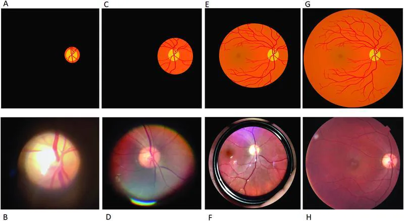
与直接检眼镜相比,放大倍率较低,但凭借宽广的视野、立体视以及与巩膜压迫的结合,能够高灵敏度地检测周边部视网膜裂孔、视网膜脱离和格子状变性。该检查是玻璃体视网膜疾病诊疗中不可或缺的检查,视网膜脱离术前全周详细眼底评估离不开BIO。美国眼科学会(AAO)2025年Preferred Practice Pattern也推荐在急性后玻璃体脱离、视网膜裂孔和格子状变性的评估中使用散瞳下BIO和巩膜压迫[2]。
Corr RH. Fundoscopy in the smartphone age: current ophthalmoscopy methods in neurology. Arq Neuropsiquiatr. 2023;81(5):502-509. Figure 4. PMCID: PMC10232018. License: CC BY.
Sen M, Honavar SG. Charles L. Schepens: Eye Spy. Indian J Ophthalmol. 2023;71(7):2625-2627. PMID: 37417098. PMCID: PMC10491037.
Kim SJ, Bailey ST, Kovach JL, et al. Posterior Vitreous Detachment, Retinal Breaks, and Lattice Degeneration Preferred Practice Pattern®. Ophthalmology. 2025;132(4):P163-P196. PMID: 39918519.
Raevis J, Hariprasad SM, Shrier E. The Depressing Part of Retina: A Review of Scleral Depression and Scleral Indentation. Ophthalmic Surg Lasers Imaging Retina. 2021;52(2):71-74. PMID: 33626165.
Lin AC, Kalaw FGP, Schönbach EM, et al. The Sensitivity of Ultra-Widefield Fundus Photography Versus Scleral Depressed Examination for Detection of Retinal Horseshoe Tears. Am J Ophthalmol. 2023;255:73-79. PMID: 37468086.
Natkunarajah M, Goldsmith C, Goble R. Diagnostic effectiveness of noncontact slitlamp examination in the identification of retinal tears. Eye (Lond). 2003;17(5):607-609. PMID: 12855967.
Trevino R, Stewart B. Change in intraocular pressure during scleral depression. J Optom. 2015;8(4):244-251. PMID: 25444648.
Dhaliwal C, Wright E, Graham C, McIntosh N, Fleck BW. Wide-field digital retinal imaging versus binocular indirect ophthalmoscopy for retinopathy of prematurity screening: a two-observer prospective, randomised comparison. Br J Ophthalmol. 2009;93(3):355-359. PMID: 19028742.
Rai AS, Rai AS, Mavrikakis E, Lam WC. Teaching binocular indirect ophthalmoscopy to novice residents using an augmented reality simulator. Can J Ophthalmol. 2017;52(5):430-434. PMID: 28985799.