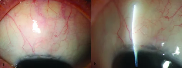
Furrer S, et al. Evaluation of filtering blebs using the ‘Wuerzburg bleb classification score’ compared to clinical findings. BMC Ophthalmol. 2012. Figure 1. PMCID: PMC3439283. License: CC BY 2.0 (https://creativecommons.org/licenses/by/2.0/).
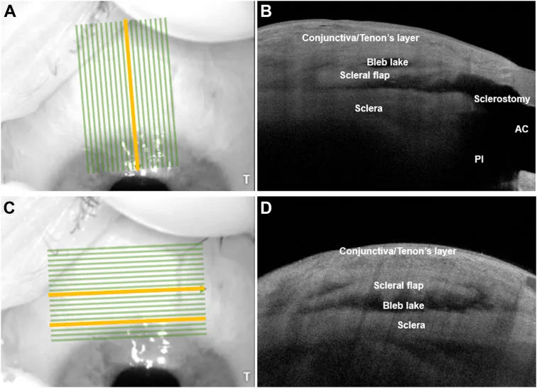
Tan JCK, Roney M, Choudhary A, Batterbury M, Vallabh NA. Visualization of Scleral Flap Patency in Glaucoma Filtering Blebs Using OCT. Ophthalmol Sci. 2025;5(1):100604. Figure 1. PMCID: PMC11483294. License: CC BY 4.0 (https://creativecommons.org/licenses/by/4.0/).
手術で作る構造を、術後の前眼部 OCT で確かめた図である。左の A と C は同じ眼を正面から撮った画像で、緑の線が OCT で走査した範囲、黄色の線がそれぞれ右の B・D の断面位置を示す。B(縦方向の断面)を上から見ると、いちばん上の層が結膜・テノン嚢(Conjunctiva/Tenon’s layer)で濾過胞の壁にあたり、その下の黒く抜けた空間が濾過胞内にたまった房水(Bleb lake)、さらにその下に基本手順3で作った半層の強膜弁(Scleral flap)、その下が強膜本体(Sclera)である。右端で強膜弁の下が前房(AC)につながっている部分が、手順5で輪部組織を切除して作った房水の出口(Sclerostomy)で、その下の PI は手順6の周辺虹彩切除部を指す。D(輪部に平行な断面)では、強膜弁の下に房水の通り道が横方向にも広がっていることが分かる。緑線・黄線と「T」の文字は撮影位置と方向を示す印であり、病変ではない。
TVT study(Tube Versus Trabeculectomy Study)の5年成績では、線維柱帯切除術とBaerveldt緑内障インプラント術の眼圧コントロールに有意差はなかった10)。ただし累積失敗率は線維柱帯切除術群(46.9%)がチューブシャント群(29.8%)より有意に高かった10)。
Fang CEH, Hakim MT, Siddiqui MM, Armstrong D, Shankar V. A retrospective study to assess visual field improvement following augmented trabeculectomy. Indian J Ophthalmol. 2023;71(5):1953-1959. doi:10.4103/ijo.IJO_2071_22.
Kandarakis SA, Karampelas M, Soumplis V, et al. Acute trabeculectomy failure following herpes zoster ophthalmicus. Ther Adv Ophthalmol. 2021;13:25158414211022461.
Barbosa RS, Salgado CM, Salgado RS, et al. Serous retinal detachment resolution after trabeculectomy in Sturge-Weber syndrome. J Glaucoma. 2021;30:e325-e328.
Chanbour W, Chanbour H, Tomey KF, Khoueir Z. Spontaneous Trabeculectomy Bleb Reformation and Regain of Function Following Phacoemulsification. Cureus. 2021;13(8):e16979. doi:10.7759/cureus.16979.
Gur Gungor S, Ceylan OM, Gul A, et al. Scleral flap wound dehiscence with Valsalva maneuver years after trabeculectomy with mitomycin C. Case Rep Ophthalmol Med. 2022;2022:8534641.
Gedde SJ, Vinod K, Wright MM, et al. Primary Open-Angle Glaucoma Preferred Practice Pattern. Ophthalmology. 2021 Jan;128(1):P71-P150. doi:10.1016/j.ophtha.2020.10.022. PMID:34933745.
Pazos M, Traverso CE, Viswanathan A; European Glaucoma Society. European Glaucoma Society - Terminology and guidelines for glaucoma, 6th Edition. Br J Ophthalmol. 2025;109(Suppl 1):1-212. doi:10.1136/bjophthalmol-2025-egsguidelines. PMID:41026937.
Yamamoto T, Sawada A, Mayama C, Araie M, Ohkubo S, Sugiyama K, et al. The 5-year incidence of bleb-related infection and its risk factors after filtering surgeries with adjunctive mitomycin C: collaborative bleb-related infection incidence and treatment study 2. Ophthalmology. 2014;121(5):1001-6. doi:10.1016/j.ophtha.2013.11.025. PMID:24424248.
Gedde SJ, Herndon LW, Brandt JD, Budenz DL, Feuer WJ, Schiffman JC, et al. Postoperative complications in the Tube Versus Trabeculectomy (TVT) study during five years of follow-up. American journal of ophthalmology. 2012;153(5):804-814.e1. doi:10.1016/j.ajo.2011.10.024. PMID:22244522; PMCID:PMC3653167.
Sugimoto Y, Mochizuki H, Ohkubo S, et al. Intraocular pressure outcomes and risk factors for failure in the collaborative bleb-related infection incidence and treatment study. Ophthalmology. 2015;122:2223-2233. doi:10.1016/j.ophtha.2015.06.038.
Kirwan JF, Lockwood AJ, Shah P, Macleod A, Broadway DC, King AJ, et al. Trabeculectomy in the 21st century: a multicenter analysis. Ophthalmology. 2013;120(12):2532-2539. doi:10.1016/j.ophtha.2013.07.049. PMID:24070811.
Morinelli EN, Sidoti PA, Heuer DK, Minckler DS, Baerveldt G, LaBree L, et al. Laser suture lysis after mitomycin C trabeculectomy. Ophthalmology. 1996;103(2):306-14. doi:10.1016/s0161-6420(96)30699-4. PMID:8594519.