Bleb-related infection (BRI) is an infectious complication that occurs after glaucomafiltration surgery (e.g., trabeculectomy) when bacteria enter through the bleb. When the infection is confined to the bleb, it is called blebitis; when it spreads into the eye, it is called bleb-associated endophthalmitis (BAE). These two conditions are managed separately in clinical practice.
Blebitis: Infection confined to the bleb or its surroundings. May be accompanied by mild to moderate anterior chamber reaction.
Bleb-associated endophthalmitis (BAE): Infection extending into the vitreous. Visual prognosis is significantly poor.
Unlike other intraocular surgeries, filtering surgery carries a risk of bleb infection not only in the early postoperative period but also over the long term. In a multicenter prospective study of trabeculectomy with mitomycin C (Collaborative Bleb-Related Infection Incidence and Treatment Study), the cumulative incidence of bleb-related infection at 5 years postoperatively was 2.2%, with bleb-associated endophthalmitis (BAE) accounting for 1.1% 5). This study is a nationwide prospective survey conducted by the Japan Glaucoma Society and provides highly reliable data. The frequency of bleb-related infection after 1 month postoperatively has been reported as 0.97–5% 4), which is higher than the incidence of late-onset infections after other intraocular surgeries. Bleb infection after glaucoma surgery often occurs several years after surgery and has a higher incidence than after cataract surgery.
Based on the time of onset, bleb-related infections are broadly classified into early-onset (within 1 month postoperatively) and late-onset (after 1 month) types 1). Early-onset infections are often caused by low-virulence organisms (coagulase-negative staphylococci), while late-onset infections involve high-virulence organisms (Streptococcus species, Haemophilus influenzae) and tend to have a poor prognosis 1). The frequency of early bleb infection is 0.1–0.2%, lower than that of late infection 4). It is important to fully inform patients who have undergone filtering surgery about the risk of late infection and instruct them to seek immediate medical attention if they experience symptoms suggestive of infection, such as redness, tearing, blurred vision, or eye pain4).
QWhat is the difference between blebitis and bleb-associated endophthalmitis?
A
Blebitis is a condition in which the infection is confined to the filtering bleb, and good visual prognosis can be expected with appropriate topical treatment. In contrast, bleb-associated endophthalmitis (BAE) is a condition in which the infection has spread to the vitreous, and even with aggressive treatment, the visual prognosis is often poor. If hypopyon and obvious bleb infection are present, it should be treated as endophthalmitis unless another cause is proven 1).
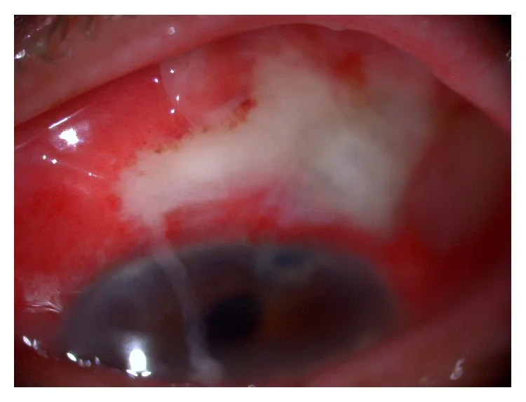
Ohtomo K, Mayama C, Ueta T, et al. Outcomes of Late-Onset Bleb-Related Endophthalmitis Treated with Pars Plana Vitrectomy. J Ophthalmol. 2015;2015:923857. Figure 1. PMID: 26495137; PMCID: PMC4606135; DOI: 10.1155/2015/923857. License: CC BY.
A characteristic “white-on-red” finding of bleb infection, where a yellow-white cloudy bleb stands out against the hyperemic conjunctiva. This corresponds to the appearance evaluation of Stages I–II discussed in the section “2. Main Symptoms and Clinical Findings.”
“White-on-red”: A yellowish-white, cloudy filtering bleb overlying a hyperemic conjunctiva is characteristic.
Bleb opacification: The bleb becomes yellowish-white and opaque.
Mild anterior chamber reaction: May or may not be accompanied by hypopyon.
Stage II-III (BAE)
Marked conjunctival hyperemia: More extensive and severe than in Stage I.
Hypopyon: Accompanied by fibrin exudation.
Vitreous opacity: Appears in Stage III. In IIIb, the fundus is not visible.
When hypopyon and obvious bleb infection are present, it should be treated as endophthalmitis unless another cause is proven1). Bleb leakage is often observed, but inflammatory debris may temporarily occlude the leakage site.
To predict filtration function after glaucoma surgery and to detect complications early, the bleb must be carefully examined at each visit. The following five items are assessed using slit-lamp microscopy.
Extent (spread): Diffuse blebs have better intraocular pressure-lowering effect than localized ones. An overhanging bleb that droops toward the cornea may interfere with vision.
Height: A low, flat bleb suggests reduced filtration function.
Wall thickness: In cases where antimetabolites are used, the bleb wall is frequently thinned. A thin-walled bleb often provides good intraocular pressure reduction but is prone to aqueous humor leakage. An encapsulated bleb, where the bleb cavity is surrounded by a thick membrane, often results in poor intraocular pressure control.
Vascularity: An avascular, ischemic bleb has a high risk of aqueous humor leakage.
Bleb leakage (greatest risk factor): Increases infection risk by 26-fold 1). Aqueous humor leakage from the bleb is a major risk factor 4)7), and late leakage carries a higher risk than early leakage 1). Oozing of aqueous humor may be observed with caution for infection, but clear leakage generally requires treatment.
Use of antimetabolites (MMC, 5-FU): Reduces goblet cells, leading to decreased mucin production and weakening of the conjunctival barrier. Promotes overall thinning and avascularity of the bleb wall; with mitomycin C use, the 5-year leakage risk reaches 15% 11).
Thin, avascular bleb wall: Common after trabeculectomy with antimetabolites. A thin-walled bleb is prone to aqueous humor leakage, which is a risk factor for bleb infection.
Inferior bleb: Increased infection rate due to exposure to tear film and lack of upper eyelid protection. Infection rate is 7.8% per patient-year for inferior blebs compared to 1.3% for superior blebs 1).
Limbus-based conjunctival incision: Higher infection rate compared to fornix-based incision (8% vs 0%) 1). Avascular blebs are more likely to occur with limbus-based technique.
Others: Conjunctivitis, blepharitis, upper respiratory tract infection, young age, axial myopia, chronic use of antibiotics.
The causative bacteria vary depending on the time of onset. Over 100 causative microorganisms have been reported in the literature 1).
Time of onset
Main causative bacteria
Characteristics
Early onset
Coagulase-negative staphylococci
Low virulence, relatively good prognosis
Late onset
Streptococcus, H. influenzae
High virulence, poor prognosis
Gram-positive cocci are the most common, with Streptococcus (approximately 385 isolates) and Staphylococcus (approximately 296 isolates) being the main ones. Among Gram-negative bacteria, Moraxella (approximately 79 cases) and Haemophilus (approximately 63 cases) are frequent 1). Rarely, zoonotic infections such as Capnocytophaga canimorsus have also been reported 2). A retrospective study in Sweden reported that the incidence of endophthalmitis and severe blebitis after trabeculectomy was 7.2 per 1000 procedures 12).
QWhat is the greatest risk factor for bleb infection?
A
Aqueous humor leakage from the filtering bleb is the greatest risk factor, increasing infection risk by 26 times 1). The use of antimetabolites thins and devascularizes the bleb wall, increasing leakage risk. Leakage is particularly likely in ischemic blebs with thin walls and poor vascularity. It is important to check for leakage using the Seidel test at every examination.
Observation of the bleb appearance is fundamental; check the five items mentioned above (extent, height, wall thickness, vascularity, and Seidel test).
The following are representative methods for classifying blebs based on appearance.
Moorfields bleb grading system (MBGS): A classification method that standardizes the evaluation of bleb morphology8)
Indiana bleb appearance grading scale (IBAGS): A classification method that systematically grades the appearance of blebs9)
Both are useful for evaluating filtration function and predicting infection risk. Combined use of anterior segment OCT allows more objective evaluation of the internal structure and wall thickness of the bleb10).
Conjunctival swab and culture: Collect specimens from purulent discharge and inoculate onto blood agar, chocolate agar, thioglycolate medium, etc.1)
Anterior chamber and vitreous tap: Essential for Stage II or higher. Perform intravitreal antibiotic injection simultaneously with culture.
PCR: Since the negative culture rate is high (21–86%), PCR is useful as a complement. Reports indicate that the detection rate improved from 47.6% to 95.3%1).
B-scan ultrasound: When the fundus is not visible in Stage IIIb, confirm vitreous opacity and choroidal thickening1).
Anterior segment OCT: Useful for evaluating the internal structure of the bleb. It allows observation of scleral flap opening, bleb cavity, microcysts, etc.10)
Endophthalmitis after cataract surgery: The onset time and surgical procedure differ. Post-cataractendophthalmitis usually occurs early (around 1 week postoperatively), while bleb-related infection often occurs several years after surgery. Differentiate by the presence or absence of bleb infection findings.
Acute anterior uveitis: The presence or absence of bleb opacity and marked surrounding injection (white-on-red) is key for differentiation. Non-infectious uveitis does not involve whitening of the bleb itself.
Encapsulated bleb: A non-infectious complication occurring several weeks after surgery. The bleb cavity is surrounded by a thick membrane, leading to poor intraocular pressure reduction, but lacks infectious findings (white-on-red).
If there is no vitreous involvement, aggressive topical antibiotic therapy is performed. If the infection is confined to the bleb and anterior chamber, it can be managed with topical and systemic antibiotics.
Intensified topical antibiotic regimen: Vancomycin (25–50 mg/mL) or cefazolin (50 mg/mL) and tobramycin (14 mg/mL) administered alternately every 30 minutes. After 48 hours, taper according to improvement.
Alternative regimen: Fourth-generation fluoroquinolone (e.g., moxifloxacin) every hour.
Subconjunctival injection: Depending on the stage of bleb infection, subconjunctival or intracameral antibiotic injection may be performed.
Consider adding topical steroids 24 hours after clinical improvement, but careful judgment is required as there are reports of worsened prognosis. Adjust antibiotics based on culture results and monitor treatment response closely. Start steroids only after confirming that the infection is controlled.
Treatment of bleb-associated endophthalmitis (BAE)
Perform intravitreal antibiotic injection simultaneously with vitreous tap for specimen collection.
Drug
Dose
Notes
Vancomycin
1 mg/0.1 mL
Gram-positive coverage
Ceftazidime
2.25 mg/0.1 mL
Gram-negative coverage
Dexamethasone
0.4 mg/0.1 mL
Adjuvant (anti-inflammatory)
When infection spreads to the vitreous cavity, vitrectomy is often required. Some reports indicate that vitrectomy (PPV) leads to better visual outcomes than needle aspiration, while recent data suggest comparable visual outcomes between the two. Since visual prognosis is extremely poor once infection reaches the vitreous cavity, prompt intervention is necessary. The EndophthalmitisVitrectomy Study (EVS) focused on post-cataract surgery endophthalmitis; patient background, causative organisms, and pathogenesis differ from BRI/BAE, so EVS results cannot be directly applied. In particular, BAE often involves late-onset, highly virulent bacteria, and more aggressive vitrectomy should be considered.
Prophylactic antibiotic administration is recommended, but there is no consensus on the specific drug, method, or duration4). In a study, participating specialists generally continued antibiotic eye drops for 1–3 months postoperatively, and no postoperative infections occurred during that period. Therefore, continuous use of antibiotics for 1–3 months after surgery is strongly recommended (strength of recommendation: strong recommendation to perform; quality of evidence: C)4).
A nationwide survey of bleb-related infections (104 eyes) comparing long-term antibiotic users and non-users showed that antibiotic use significantly delayed the onset of bleb infection6).
Non-use group: median time to infection 3.9 years
Long-term use group: median time to infection 6.4 years
Ointment group: median time to infection 10.5 years
If the bleb wall is thin, with aqueous humor oozing upon eyelid elevation, or if the patient reports tearing upon waking, consider instilling a new quinolone antibiotic ophthalmic ointment before bedtime4).
Resistant bacteria were found in 9 of 26 eyes in the long-term use group, but 6 of those were Staphylococcus epidermidis, which is less likely to cause severe endophthalmitis4). Indiscriminate use of antibiotics should also be avoided from a health economic perspective.
Autologous blood injection: Injection of autologous blood into and around the filtering bleb. May cause a sudden increase in intraocular pressure.
Surgical treatment: Conjunctival advancement (success rate 100%), amniotic membrane transplantation (success rate 45%). If the risk of infection is high or hypotony does not resolve, bleb reconstruction with a free conjunctival graft or a pedicled conjunctival flap from above is indicated. Long-term reassessment of bleb morphology and intraocular pressure management should be performed concurrently 11)
QHow long should antibiotics be used after glaucoma surgery?
A
The Glaucoma Practice Guidelines (5th edition) strongly recommend continued use of antibiotics for 1 to 3 months postoperatively (strength of evidence C) 4). Thereafter, depending on the risk of infection such as the presence of bleb leak, long-term use of a new quinolone ophthalmic ointment before bedtime should be considered. There is a report that long-term use significantly delays the median onset of infection from 3.9 years in the non-use group to 10.5 years in the ointment group 6). However, indiscriminate use should be avoided, and the decision to continue should be made in consultation with the attending physician.
6. Pathophysiology and Detailed Mechanism of Onset
The pathology of filtering bleb-related infections stems from the filtering bleb formed by glaucomafiltration surgery serving as the portal of entry for infection.
In trabeculectomy, aqueous humor is directed from under the scleral flap to the subconjunctival space to form a filtering bleb. If the bleb wall becomes thin or leaks, tears or periocular commensal bacteria can enter the eye.
The use of antimetabolites (MMC, 5-FU) increases infection risk through the following combined mechanisms:
Reduces conjunctival goblet cell count, decreasing mucin production and impairing tear film defense function
Promotes generalized thinning and avascularity of the conjunctiva, weakening the mechanical strength of the bleb wall
Weakens physical and immunological barriers, facilitating bacterial entry from the conjunctival surface into the eye
Antimetabolites are essential for preventing scarring in current trabeculectomy, but the long-term increased infection risk associated with their use must always be kept in mind.
Bleb morphology changes over time. In trabeculectomy, scar tissue forms in the subconjunctival space during postoperative wound healing. Concomitant use of mitomycin C (MMC) suppresses excessive early tissue reaction and increases the likelihood of long-term bleb maintenance, but outflow resistance of the scleral flap and bleb morphology change rapidly during the first few months after surgery when scarring progresses quickly. Without appropriate management during this period, bleb wall thinning is likely to progress.
Avascular ischemic blebs have a particularly high risk of aqueous humor leakage. Avascular blebs are more likely to occur with limbal-based conjunctival incisions, which is also related to the higher infection rate of limbal-based incisions (8% vs. 0% for fornix-based incisions) 1). Aqueous humor leakage (oozing or frank leakage) and bleb infection, where bacteria enter the bleb, are long-term complications requiring attention. Oozing may be observed with caution for infection, but frank leakage is an indication for intervention.
Relationship between causative organisms and disease stage
The pathogenicity of causative organisms correlates with the time of onset 1). Early-onset cases are predominantly caused by low-virulence bacteria (coagulase-negative staphylococci), similar to endophthalmitis after cataract surgery, originating from the normal flora of tears and eyelids. These bacteria do not produce exotoxins, and appropriate treatment leads to a good prognosis.
In contrast, late-onset cases involve more virulent bacteria such as Streptococcus species (S. pneumoniae, S. viridans group), Haemophilus influenzae, and Serratia species 1). These bacteria have high exotoxin production and tissue invasiveness, leading to rapid clinical progression and poor visual prognosis.
Kandarakis et al. (2022) comprehensively reviewed BRI causative organisms reported in the literature and identified over 100 microorganisms. Among Gram-positive cocci, Streptococcus (approximately 385 isolates) and Staphylococcus (approximately 296 isolates) were overwhelmingly predominant, while among Gram-negative bacteria, Moraxella (approximately 79 cases) and Haemophilus (approximately 63 cases) were the main ones1).
Yang et al. (2021) reported an 81-year-old man who developed blebitis caused by Capnocytophaga canimorsus 10 years after trabeculectomy. The patient’s face was regularly licked by his pet dog, and it was thought that the dog’s oral commensal bacteria infected the filtering bleb. Good visual prognosis (20/70) was achieved with trabeculectomy revision2).
7. Latest Research and Future Perspectives (Investigational Reports)
The review by Kandarakis et al. (2022) points out the problem of high negative culture rates of 21–86% with conventional methods. Real-time PCR improves bacterial detection rates from 47.6% to 95.3%. Furthermore, comprehensive microbial identification by metagenomics (high-throughput DNA sequencing) is expected as a next-generation diagnostic method1).
Reduction of Infection Risk with Minimally Invasive Glaucoma Surgery (MIGS)
Reviews of MIGS indicate that it is expected to reduce invasiveness and decrease serious postoperative complications compared to conventional filtering surgery. However, some procedures involve devices that form filtering blebs, and infection risk must be evaluated for each type of surgery3).
The report of Capnocytophaga canimorsus blebitis by Yang et al. (2021) showed that contact with pet animals can be a new risk factor for infection in eyes with a history of filtering surgery. DNA sequencing has enabled identification of pathogens that were previously difficult to culture2).
The Glaucoma Practice Guidelines (5th edition) state that while long-term postoperative antibiotic use significantly delays the onset of infection, there is no consensus on the drug, method, or duration of administration, and high-evidence-level studies are awaited4). Along with changes in conjunctival flora and the emergence of resistant bacteria, long-term RCTs are an important future issue.
Kandarakis SA, Doumazos L, Mitsopoulou D, Economou MA, Mylona I, Dimitriou C, et al. A Review on Pathogens and Necessary Diagnostic Work for Bleb-Related Infections (BRIs). Diagnostics (Basel, Switzerland). 2022;12(9). doi:10.3390/diagnostics12092075. PMID:36140477; PMCID:PMC9497804.
Yang MC, Ling J, Mosaed S. Capnocytophaga canimorsus blebitis: case report and review of literature. BMC ophthalmology. 2021;21(1):59. doi:10.1186/s12886-021-01823-8. PMID:33499831; PMCID:PMC7839216.
Balas M, Mathew DJ, Bicket AK. Minimally Invasive Glaucoma Surgery: A Review of the Literature. Vision (Basel). 2023;7(3):54. doi:10.3390/vision7030054. PMID: 37606500.
Yamamoto T, Sawada A, Mayama C, et al. The 5-year incidence of bleb-related infection and its risk factors after filtering surgeries with adjunctive mitomycin C: collaborative bleb-related infection incidence and treatment study 2. Ophthalmology. 2014;121(5):1001-1006. doi:10.1016/j.ophtha.2013.11.025. PMID: 24424248.
Sagara H, Yamamoto T, Imaizumi K, Sekiryu T. Impact of topically administered steroids, antibiotics, and sodium hyaluronate on bleb-related infection onset: the Japan Glaucoma Society survey of bleb-related infection report 4. J Ophthalmol. 2017;2017:7062565. doi:10.1155/2017/7062565. PMID: 29138694. PMCID: PMC5613473.
Yamamoto T, Kuwayama Y, Kano K, et al. Clinical features of bleb-related infection: a 5-year survey in Japan. Acta Ophthalmol. 2013;91(7):619-624. doi:10.1111/j.1755-3768.2012.02480.x. PMID: 22883301.
Wells AP, Ashraff NN, Hall RC, et al. Comparison of two clinical bleb grading systems. Ophthalmology. 2006;113(1):77-83. doi:10.1016/j.ophtha.2005.06.037. PMID: 16389104.
Cantor LB, Mantravadi A, WuDunn D, Swamynathan K, Cortes A. Morphologic classification of filtering blebs after glaucomafiltration surgery: the Indiana Bleb Appearance Grading Scale. J Glaucoma. 2003;12(3):266-71. doi:10.1097/00061198-200306000-00015. PMID:12782847.
Kojima S, Inoue T, Kawaji T, Tanihara H. Filtration bleb revision guided by 3-dimensional anterior segment optical coherence tomography. J Glaucoma. 2014;23(5):312-315. doi:10.1097/IJG.0b013e3182741ee6. PMID: 23377583.
Kim EA, Law SK, Coleman AL, et al. Long-Term Bleb-Related Infections After Trabeculectomy: Incidence, Risk Factors, and Influence of Bleb Revision. Am J Ophthalmol. 2015;159(6):1082-1091. doi:10.1016/j.ajo.2015.03.001. PMID: 25748577.
Wallin Ö, Al-ahramy AM, Lundström M, Montan P. Endophthalmitis and severe blebitis following trabeculectomy: epidemiology and risk factors; a single-centre retrospective study. Acta Ophthalmol. 2014;92(5):426-431. doi:10.1111/aos.12257. PMID: 24020653.
Copy the article text and paste it into your preferred AI assistant.
Article copied to clipboard
Open an AI assistant below and paste the copied text into the chat box.