
Lacrimal irrigation test (Lacrimal Irrigation Test)
1. What is lacrimal irrigation testing
Section titled “1. What is lacrimal irrigation testing”Lacrimal irrigation test (lacrimal irrigation test / lacrimal syringing) is a functional test in which saline is injected from the punctum with a syringe attached to a lacrimal irrigation needle to infer whether there is a passage obstruction and the site of obstruction in the lacrimal drainage system. By observing whether the solution flows into the nasal cavity and pharynx (the patient’s sensation), whether there is reflux from the punctum, and the character of the reflux fluid (serous or purulent), the presence and site of obstruction are assessed.
After topical anesthesia, inserting while keeping the anatomy of the vertical and horizontal parts of the canaliculus in mind leads to a more accurate assessment. The procedure is simple and widely used as an outpatient procedure, and it is regarded as an essential test in the initial evaluation of patients who complain of epiphora and eye discharge.
Indications
Section titled “Indications”Lacrimal irrigation is actively performed in the following situations. Because lacrimal duct obstruction is often hidden behind recurrent conjunctivitis and keratitis, it is recommended to perform it proactively in routine practice to avoid missing it.
- Epiphora — the most common chief complaint
- Chronic or recurrent eye discharge
- Recurrent conjunctivitis or keratitis (to rule out lacrimal duct obstruction)
- Suspected dacryocystitis (swelling over the lacrimal sac and redness at the inner corner of the eye)
- Confirmation of patency after removal of a lacrimal tube
It is performed in patients with epiphora (tearing), chronic eye discharge (eye mucus), and recurrent conjunctivitis or keratitis. If lacrimal duct obstruction is missed, it can cause chronic infection, so it is important to actively perform the test in these symptoms. It is also used to confirm patency after removal of a lacrimal tube.
2. Pre-examination checks
Section titled “2. Pre-examination checks”Checking medication use history
Section titled “Checking medication use history”The following medications can cause lacrimal duct disorders and must be checked before the examination.
- Fluoropyrimidine anticancer drugs, including TS-1 (tegafur/gimeracil/oteracil potassium): they can cause lacrimal duct obstruction. If the blockage progresses, treatment becomes difficult, so if you feel strong resistance when inserting the irrigation cannula, place a lacrimal tube early to help prevent the obstruction from worsening.
- Rebamipide eye drops (rebamipide): used for dry eye, but they can harden inside the lacrimal drainage system and cause dacryolithiasis.
Confirm anterior segment findings
Section titled “Confirm anterior segment findings”Evaluate the following before irrigation testing.
- Tear fluid characteristics: serous and clear is normal. Viscous or purulent fluid suggests dacryocystitis or lacrimal duct obstruction.
- Krehbiel flow: observe with a slit-lamp microscope how the tear fluid flows from the punctum into the canaliculus after blinking. If the lacrimal drainage system is obstructed, absorption does not occur.
- Punctal morphology: punctal stenosis or obstruction can be caused by infection, chronic conjunctivitis, allergy, after cataract surgery, or glaucoma eye drops.
- Canaliculitis: this is especially easy to miss, and should be actively suspected if discharge is seen when the punctum is pressed.
- Dry eye, conjunctivochalasis, and eyelid abnormalities: check these to differentiate them from reflex tearing or impaired tear drainage.
3. Examination procedure (position, instruments, technique)
Section titled “3. Examination procedure (position, instruments, technique)”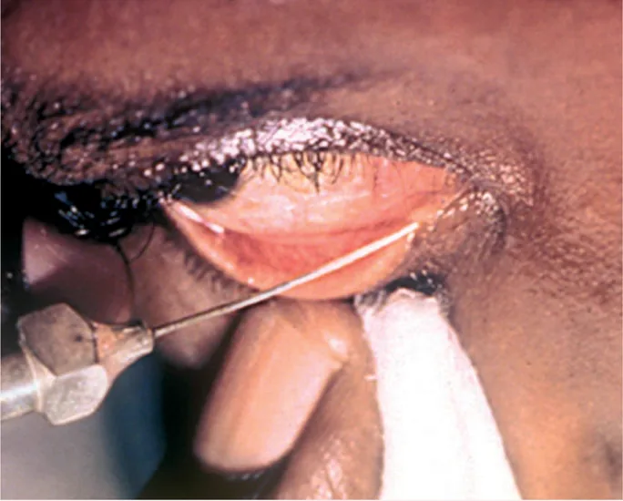
Positioning and setup
Section titled “Positioning and setup”Place the patient supine on the procedure bed. Supine positioning stabilizes the head, makes the procedure easier, and allows easier observation. Placing cut cotton on the skin outside the treated eye is convenient because it saves the trouble of repeatedly wiping away any irrigation fluid that flows back.
Having staff prepare the following in advance can shorten the procedure time:
- Placing cut cotton pads
- Topical anesthesia with 0.4% oxybuprocaine eye drops (this is sufficient in many cases)
Choosing the lacrimal irrigation needle
Section titled “Choosing the lacrimal irrigation needle”| Type | Features | Recommendation |
|---|---|---|
| Straight type | Can be inserted as far as the nasolacrimal duct | Preferably avoided by anyone other than experienced operators (risk of blind insertion) |
| Curved type | About 8 mm from the tip to the bend | Recommended for routine cases |
Irrigation fluid is often normal saline. If a mixture with povidone-iodine (16-fold dilution) is used, an added antiseptic effect can be expected. A 2.5 mL syringe is easier to handle and lets you sense resistance more clearly (compared with 5 mL).
Tips for the procedure (numbered steps)
Section titled “Tips for the procedure (numbered steps)”- Identification of the punctum: Check the upper and lower puncta; usually perform the procedure from the lower punctum
- Lid retraction: With the hand opposite the syringe, firmly pull the eyelid outward to straighten the canaliculus. If you advance the needle before it is fully straightened, the tip may touch the side wall of the canaliculus and be mistaken for blockage
- Insertion of the irrigation needle: The canaliculus runs vertically for about 2 mm from the punctum, then curves at nearly a right angle toward the lacrimal sac. Insert the irrigation needle with this anatomy in mind
- Advance to the bend: With a curved-type irrigation needle, advance it to the bend from the tip (about 8 mm) so that it reaches the common canaliculus just before the lacrimal sac
- Pressurize and assess: Gradually increase pressure as you push the syringe. Assess whether there is resistance, whether there is reflux and from which punctum the reflux occurs (same side or opposite side), and the nature of the refluxed fluid (serous or purulent)
- Confirm with the patient: Confirm whether the patient felt the irrigating fluid flow into the nasal cavity or pharynx
Because topical anesthesia with 0.4% oxybuprocaine eye drops is used, discomfort during the procedure is minimal in most cases. You may feel a slight sense of pressure when the lacrimal irrigation needle is inserted, but severe pain usually does not occur. It is important to provide enough topical anesthesia before the examination.
4. Interpretation of Results
Section titled “4. Interpretation of Results”Based on the irrigation findings, the site of obstruction is estimated in the following five patterns.
| Findings | Interpretation |
|---|---|
| Flow into the nasal cavity and pharynx (noticed by the patient) | Good irrigation flow (normal) |
| No resistance; reflux from the same-side punctum | Communication between the upper and lower lacrimal passages; obstruction distal to the common canaliculus (lacrimal sac or nasolacrimal duct) |
| Strong resistance; reflux from the same-side punctum | Obstruction of the canaliculus or common canaliculus |
| Purulent reflux fluid | Nasolacrimal duct obstruction with dacryocystitis (chronic dacryocystitis) |
| Regurgitation from the opposite punctum | Lacrimal sac/nasolacrimal duct obstruction; upper and lower canaliculi patent |
The probability that the estimated obstruction site from irrigation testing matches the actual findings during lacrimal endoscopic surgery is around 70%, so it is by no means high1). For a definitive diagnosis of the obstruction site, lacrimal endoscopy may be necessary.
5. Auxiliary tests
Section titled “5. Auxiliary tests”Dye retention test
Section titled “Dye retention test”A useful adjunctive test when lacrimal irrigation is difficult because body movement cannot be controlled, such as in children.
- Fifteen minutes after fluorescein staining, assess whether fluorescent dye remains in the conjunctival sac or flows onto the eyelids
- Its sensitivity for congenital nasolacrimal duct obstruction is said to be about 95%
- In adults, it can also be used to diagnose functional epiphora when combined with irrigation testing
Micro-Reflux test
Section titled “Micro-Reflux test”A diagnostic adjunct used when dacryocystitis is suspected. If pus refluxes when the lacrimal sac area is pressed, dacryocystitis can be diagnosed. It is also a method for simultaneously diagnosing lacrimal duct obstruction using fluorescein dye.
Dacryocystography
Section titled “Dacryocystography”After confirming obstruction with an irrigation test, it is used for a detailed anatomical assessment of the site of obstruction. It combines contrast injection and imaging, but it can sometimes be difficult to determine where the contrast has reached, and it may be combined with lacrimal endoscopy1).
6. Role of lacrimal irrigation in congenital nasolacrimal duct obstruction
Section titled “6. Role of lacrimal irrigation in congenital nasolacrimal duct obstruction”Congenital nasolacrimal duct obstruction is the most common cause of tearing and eye discharge in infants, and the irrigation test is used to decide whether to move from conservative treatment to surgery2).
The recommendations in the congenital nasolacrimal duct obstruction treatment guideline2) are as follows.
- CQ1 Lacrimal sac massage (Crigler method): Weakly recommend performing it. It is done with the aim of opening the obstructing membrane at the lower end of the nasolacrimal duct by increasing pressure
- CQ2 Topical antibiotics: Weakly recommend using them only when necessary. Use them only when signs of infection such as eye discharge or conjunctival redness are present
- CQ3 Surgical intervention (probing): In unilateral congenital nasolacrimal duct obstruction, probing under local anesthesia around 6 to 9 months of age is weakly recommended. Because the spontaneous resolution rate within the first year is high, observation until then is the rule
- Repeat probing after initial probing failure: Weakly recommend against it. In failed cases, consider lacrimal tube insertion under lacrimal endoscopy
The irrigation test is used to confirm obstruction and to decide whether surgery is appropriate between 6 and 15 months of age.
About 90% are said to resolve on their own within the first year of life, so conservative treatment (lacrimal sac massage and antibiotic eye drops) is tried first. If it is on one side and there is no improvement after 6 to 9 months of age, probing under local anesthesia is chosen. For cases on both sides or complex cases, endoscopic lacrimal surgery under general anesthesia is considered2).
7. Management of special cases
Section titled “7. Management of special cases”Lacrimal irrigation for bacterial clearance (palliative treatment)
Section titled “Lacrimal irrigation for bacterial clearance (palliative treatment)”For nasolacrimal duct obstruction with dacryocystitis, radical treatment such as lacrimal tube insertion or dacryocystorhinostomy (DCR) is appropriate. However, when surgery is difficult (poor general condition, patient refusal, etc.), palliative drainage may be done by regular lacrimal irrigation. It is important to repeat irrigation until the refluxed irrigating fluid becomes clear. A method of instilling antibiotic eye ointment into the lacrimal passage after irrigation is also used.
Anticancer drug-related lacrimal duct obstruction
Section titled “Anticancer drug-related lacrimal duct obstruction”Fluoropyrimidine anticancer drugs, including TS-1 (tegafur, gimeracil, and oteracil potassium), are known to cause lacrimal duct obstruction. If there is strong resistance when inserting the lacrimal irrigation needle, early placement of a lacrimal tube can help prevent progression of the obstruction. In patients receiving TS-1, regular lacrimal irrigation and monitoring are desirable.
Dacryolithiasis associated with Levamid eye drops
Section titled “Dacryolithiasis associated with Levamid eye drops”Levamid sodium (Levamid) eye drops, used for dry eye, can solidify within the lacrimal passages and cause dacryolithiasis. In patients using Levamid eye drops who complain of tearing and eye discharge, perform lacrimal irrigation with the possibility of dacryoliths in mind. If strong resistance is felt, confirmation with lacrimal endoscopy is useful.
Evaluation after removal of the lacrimal tube
Section titled “Evaluation after removal of the lacrimal tube”Perform lacrimal irrigation before and after removal of the lacrimal tube placed after surgery to confirm patency. Because some cases develop re-obstruction after removal, regular lacrimal irrigation during the 1 to 3 months of follow-up after removal is recommended.
8. References
Section titled “8. References”- 日本涙道・涙液学会涙道内視鏡診療の手引き作成委員会. 涙道内視鏡診療の手引き. 日本眼科学会雑誌. 2023;127(10):896-917.
- 先天鼻涙管閉塞診療ガイドライン作成委員会. 先天鼻涙管閉塞診療ガイドライン. 日本眼科学会雑誌. 2022;126(11):991-1021.