The iris is a membrane-like tissue that separates the anterior and posterior chambers. The iris root is the thinnest part of the iris and is prone to rupture from trauma. When blunt trauma increases intraocular pressure, the eyeball wall deforms, applying stretching forces to the iris. If the iris root ruptures, iridodialysis occurs, and the pupil becomes displaced.
If a cleft forms slightly more scleral than the iris root within the ciliary body, it becomes angle recession. Further scleral detachment of the ciliary body from the sclera results in cyclodialysis. In penetrating trauma, the iris may prolapse through the corneoscleral laceration.
Damage to the pupillary sphincter causes traumatic mydriasis. The light reflex is diminished or absent. For these iris injuries, if symptoms persist, repair is performed via iris suturing and pupilloplasty.
QCan iris trauma be left untreated?
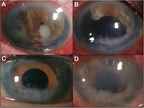
A
If the area of iris dialysis is small and there are no subjective symptoms, observation alone is often acceptable. However, if the dialysis is extensive with pupillary deviation, or if there is monocular diplopia or photophobia, surgical repair is necessary. Additionally, since anterior chamber hemorrhage or angle recession often coexists, long-term monitoring of intraocular pressure is important.
Kevin C Firl, Sandra R Montezuma. Chronic post-operative iris prosthesis endophthalmitis in a patient with traumatic aniridia: a case report. BMC Ophthalmol. 2016 Nov 9;16:197. Figure 1. PMCID: PMC5103383. License: CC BY.
Anterior segment photograph showing a large defect in iris tissue and marked deformation around the pupil. This indicates loss of iris support structures after trauma, allowing assessment of the anatomical damage that is a candidate for iris reconstruction.
Photophobia occurs when excessive light enters the eye due to increased or displaced pupil size. Monocular diplopia results from abnormal light pathways through the iris defect. Decreased visual acuity is often caused by increased higher-order aberrations or concurrent traumatic cataract/vitreous hemorrhage.
In iridodialysis, a crescent-shaped or semilunar cleft is observed with a slit-lamp microscope. Vitreous prolapse due to zonular rupture may also be present. If pupillary deviation is mild and hyphema is present after blunt trauma, suspect angle recession or cyclodialysis. In penetrating trauma, if iris incarceration or prolapse is present at the laceration site, the pupil deviates toward that direction.
Blunt trauma is the most common cause. This includes balls from ball sports, fists, branches, and flying objects. In penetrating trauma, iris prolapse occurs with corneoscleral lacerations.
Iris injury during cataract surgery is also a cause 1). Small pupil is the most important risk factor for intraoperative iris injury 1). Systemic administration of alpha-1 adrenergic receptor antagonists causes intraoperative floppy iris syndrome (IFIS), increasing the risk of iris prolapse 1). The frequency of IFIS or iris prolapse is reported as 0.5–2.0%, and iris/ciliary body injury as 0.6–1.2% 1).
Iridodialysis: Observed as a crescent-shaped iris defect. Transillumination can detect a defect in light transmission at the iris root 2).
Pupil Evaluation: Record pupil diameter, shape, and direction and degree of deviation. Check for the presence of light reflex and response to mydriatics.
Associated Injuries: Evaluate for lens subluxation, anterior capsule rupture, and vitreous prolapse.
Gonioscopy and Imaging
Gonioscopy: Assess increased distance from the iris root to the scleral spur and widening of the ciliary body band 2). Comparison with the fellow eye is important. Avoid in the presence of hyphema due to risk of rebleeding.
First, prioritize conservative treatment for hyphema. Rest and instillation of mydriatics (atropine) promote absorption of hyphema. If the iridodialysis is small and asymptomatic, surgery is not required.
Indications for Iris Suturing and Preoperative Evaluation
Surgery is indicated when there is significant pupillary deviation causing decreased visual acuity, photophobia, or monocular diplopia. The larger the pupil diameter, the greater the higher-order aberrations; a diameter exceeding 4 mm is considered to significantly reduce visual acuity. If, after IOL insertion, a mydriatic pupil of approximately 6 mm or more remains with miotics and iris traction, concurrent pupilloplasty is performed.
Preoperatively, the iris is stretched with forceps in the anterior chamber to simulate the suture position. This maneuver itself also has a miotic effect.
Modified Siepser Sliding Knot method: A curved needle is inserted through a side port, the iris is grasped 2–3 mm wide, and the needle is brought out through the opposite side port. The thread pulled out is looped and brought out through the initial side port, then tied using the Siepser slipknot technique. 9-0 or 10-0 Prolene suture is used.
Single-pass Four-throw (SFT) method: The other thread end is passed through the loop of the thread pulled out from the side port four times and tied. This method has the advantage of requiring only one intra- and extra-anterior chamber knot-tying maneuver.
McCannel method: A long needle is inserted through the main incision, and both iris edges and the limbus are pierced in one pass. Both thread ends are pulled out and tied outside the eye as in standard suturing. The advantage is that the knot-tying technique is easy to understand.
Iris cerclage: A method of continuous suturing around the iris for extensive iris dialysis. The intra-anterior chamber manipulation is complex and technically demanding.
Pick-up needle (30-gauge thin-wall needle) technique: Used when threading with a long needle is difficult. The iris is pierced from the cornea with a pick-up needle, which is then locked with the long needle and pulled out. This allows accurate threading to the target position.
QHow long does iris suture surgery take?
A
The iris suturing procedure itself is often completed within several tens of minutes. If performed simultaneously with cataract surgery or IOL insertion, additional time is added. The surgical time varies depending on the technique and the extent of iris damage.
QHow much does vision recover after surgery?
A
If the iris trauma is isolated, pupilloplasty often improves photophobia and visual acuity. There is a report of a case achieving a corrected visual acuity of 1.2 after iris suturing and IOL insertion following ocular contusion. However, if there is concomitant retinal damage or optic nerve injury, vision improvement from iris repair alone is limited.
When blunt force is applied to the eye, the intraocular pressure rises sharply. The eyeball wall deforms, and stretching and shearing forces act on the iris and ciliary body attached to the inner side. The iris root is the thinnest part of the iris and is most susceptible to damage.
If the iris root is torn, it results in iridodialysis. If the tear occurs between the circular (Müller) and longitudinal (Brücke) muscles of the ciliary body, it results in angle recession. Angle recession indicates that the ciliary body has moved posteriorly along with the iris. Furthermore, if the ciliary body detaches from the sclera on the scleral side, it results in cyclodialysis, creating an outflow pathway for aqueous humor and leading to hypotony.
Damage to the pupillary sphincter is a direct cause of traumatic mydriasis. Even minor tissue damage disrupts the blood-aqueous barrier, causing inflammatory cells to migrate into the anterior chamber (traumatic iritis). Hyphema results from vascular damage at the angle, and the breakdown products of red blood cells can obstruct aqueous outflow, causing transient intraocular pressure elevation (ghost cell glaucoma).
Intraocular iris suturing using an ultra-small curved needle (1.5 mm) and a dedicated needle holder has been reported. Compared to conventional extraocular manipulation with a long needle, it allows more precise suturing and less iris damage. Even if the needle stands vertically in the anterior chamber, it has the advantage of minimizing damage to the corneal endothelium.
Artificial iris is expected as a treatment option for cases with extensive iris damage or those unsuitable for iris suturing. It has accumulated clinical experience abroad, but is currently not approved in Japan. Iris-fixated IOLs are also being developed and may become a new option for aniridia or severe iris damage.
Pupilloplasty by iris suturing is a useful method, but it is important to handle the iris properly and accurately assess the indication. For extensive damage, artificial iris should be considered.
ESCRS. ESCRS Clinical Guidelines for Cataract Surgery. European Society of Cataract and Refractive Surgeons. 2024.
European Glaucoma Society. European Glaucoma Society Terminology and Guidelines for Glaucoma, 5th Edition. Br J Ophthalmol. 2021 Jun;105(Suppl 1):1-169. doi:10.1136/bjophthalmol-2021-egsguidelines. PMID:34675001.
Copy the article text and paste it into your preferred AI assistant.
Article copied to clipboard
Open an AI assistant below and paste the copied text into the chat box.