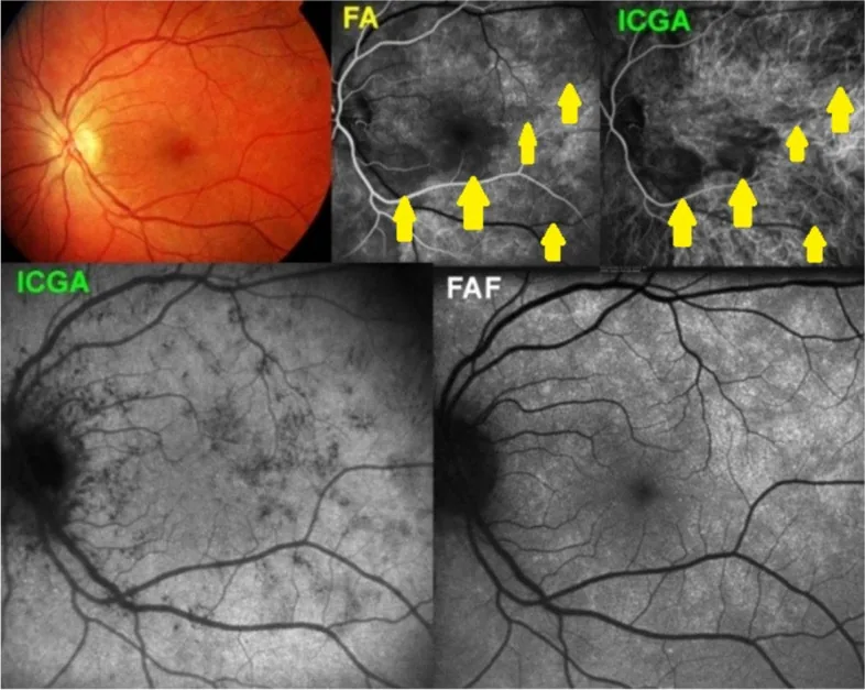
Papasavvas I, et al. Choroidal vasculitis as a biomarker of inflammation of the choroid. Indocyanine Green Angiography (ICGA) spearheading for diagnosis and follow-up, an imaging tutorial. J Ophthalmic Inflamm Infect. 2024. Figure 5. PMCID: PMC11618284. License: CC BY 4.0.
การตรวจ OCT พบว่าในระยะ active ellipsoid zone (รอยต่อระหว่าง inner segment และ outer segment ของเซลล์รับแสง) มีลักษณะไม่สม่ำเสมอและหายไป มีรายงานว่าการมองเห็นจะดีขึ้นเมื่อความผิดปกตินี้ดีขึ้น ซึ่งแสดงให้เห็นว่าความเสียหายต่อเรตินาชั้นนอกที่เกิดขึ้นชั่วคราวและสามารถกลับคืนได้นั้นเป็นลักษณะสำคัญของ MEWDS3) การตรวจ en face swept-source OCT ในปัจจุบันสามารถมองเห็นขอบเขตของรอยโรคที่ยากต่อการตรวจพบด้วยการตรวจตัดขวางแบบเดิมได้ 3)
OCT แบบ en face swept-source : มีรายงานเพิ่มขึ้นเรื่อยๆ ว่าเป็นวิธีการวินิจฉัยใหม่ที่สามารถมองเห็นรูปร่าง การกระจาย และขอบเขตชั้นในของรอยโรคจุดขาวได้ในสามมิติ3)
Testi I, Modugno RL, Pavesio C. Multimodal imaging supporting the pathophysiology of white dot syndromes. Journal of ophthalmic inflammation and infection. 2021;11(1):32. doi:10.1186/s12348-021-00261-3. PMID:34529201; PMCID:PMC8446150.
Pichi F, Srvivastava SK, Chexal S, Lembo A, Lima LH, Neri P, et al. EN FACE OPTICAL COHERENCE TOMOGRAPHY AND OPTICAL COHERENCE TOMOGRAPHY ANGIOGRAPHY OF MULTIPLE EVANESCENT WHITE DOT SYNDROME: New Insights Into Pathogenesis. Retina. 2016;36(Supplement 1):S178-S188. doi:10.1097/IAE.0000000000001255. PMID:28005676.
Yannuzzi NA, Swaminathan SS, Zheng F, Miller A, Gregori G, Davis JL, Rosenfeld PJ.. Swept-Source OCT Angiography Shows Sparing of the Choriocapillaris in Multiple Evanescent White Dot Syndrome. Ophthalmic Surg Lasers Imaging Retina. 2017;48(1):69-74. doi:10.3928/23258160-20161219-10. PMID:28060397.
Mantovani A, Invernizzi A, Staurenghi G, Herbort CP Jr. Multiple Evanescent White Dot Syndrome: A Multimodal Imaging Study of Foveal Granularity. Ocular immunology and inflammation. 2019;27(1):141-147. doi:10.1080/09273948.2017.1353104. PMID:28981397.
Dell’Omo R, Wong R, Marino M, Konstantopoulou K, Pavesio C. Relationship between different fluorescein and indocyanine green angiography features in multiple evanescent white dot syndrome. British Journal of Ophthalmology. 2010;94(1):59-63. doi:10.1136/bjo.2009.163550. PMID:19692364.
Jampol LM, Becker KG. White spot syndromes of the retina: a hypothesis based on the common genetic hypothesis of autoimmune/inflammatory disease. American journal of ophthalmology. 2003;135(3):376-9. doi:10.1016/s0002-9394(02)02088-3. PMID:12614757.
Khochtali S, Dridi T, Abroug N, Ksiaa I, Lupidi M, Khairallah M. Swept-Source Optical Coherence Tomography Angiography Shows Choriocapillaris Flow Reduction in Multiple Evanescent White Dot Syndrome. Journal of current ophthalmology. 2020;32(2):211-215. doi:10.4103/JOCO.JOCO_107_20. PMID:32671309; PMCID:PMC7337020.
Zicarelli F, Mantovani A, Preziosa C, Staurenghi G. Multimodal Imaging of Multiple Evanescent White Dot Syndrome: A New Interpretation. Ocul Immunol Inflamm. 2020;28(5):814-820. doi:10.1080/09273948.2019.1635169. PMID:31414928.
Wang L, Lankry P, Rabinovitch D, Gallo R, Laiginhas R, Iyer P, Shulman S, Trivizki O.. An Elderly Man with Atypical Multiple Evanescent White Dot Syndrome. Case Rep Ophthalmol. 2022;13(3):892-896. doi:10.1159/000526583. PMID:36466057; PMCID:PMC9710463.
Ramirez Marquez E, Ayala Rodríguez SC, Rivera L, Pappaterra-Rodriguez MC, Requejo-Figueroa GA, Rios R, Rivera-Grana E, Rodríguez-García EJ, Oliver AL.. Contralateral Recurrences of Post-vaccination Multiple Evanescent White Dot Syndrome. Cureus. 2022;14(12):e32300. doi:10.7759/cureus.32300. PMID:36628035; PMCID:PMC9822784.
Smeller L, Toth-Molnar E, Sohar N.. White Dot Syndrome Report in a SARS-CoV-2 Patient. Case Rep Ophthalmol. 2022;13(3):744-750. doi:10.1159/000526090. PMID:36845457; PMCID:PMC9944209.
Shepherd EA, Herst BS, Parlitsis GJ, Shapiro MJ, Goldstein DA, Merrill PT.. Multiple Evanescent White-Dot Syndrome in a 9-Year-Old Girl. J Vitreoretin Dis. 2022;6(6):470-473. doi:10.1177/24741264211044222. PMID:37009534; PMCID:PMC9954785.
Chen N, Mandell M, Arjmand P. Multimodal imaging findings of multiple evanescent white dot syndrome in COVID-19 patients. IDCases. 2024;38:e02110. doi:10.1016/j.idcr.2024.e02110. PMID:39582747; PMCID:PMC11585666.
Yasuda E, Matsumiya W, Maeda Y, Kusuhara S, Nguyen QD, Nakamura M, Hara R.. Multiple evanescent white dot syndrome following BNT162b2 mRNA COVID-19 vaccination. Am J Ophthalmol Case Rep. 2022;26:101532. doi:10.1016/j.ajoc.2022.101532. PMID:35434421; PMCID:PMC8994682.
Wiley ZC, Pakravan M, Charoenkijkajorn C, et al. Uveomeningeal syndrome presenting with bilateral optic disc edema and multiple evanescent white dots syndrome (MEWDS). Am J Ophthalmol Case Rep. 2022;26:101538. doi:10.1016/j.ajoc.2022.101538. PMID:35514800; PMCID:PMC9062123.
Jampol LM, Sieving PA, Pugh D, Fishman GA, Gilbert H. Multiple evanescent white dot syndrome. I. Clinical findings. Arch Ophthalmol. 1984;102(5):671-674. doi:10.1001/archopht.1984.01040030527008. PMID:6721749.
Finn AP, Khurana RN. Multiple evanescent white dot syndrome: Bilateral disease may be silent and asymmetric. Am J Ophthalmol Case Rep. 2021;21:101004. doi:10.1016/j.ajoc.2020.101004. PMID:33376834; PMCID:PMC7758516.
Figueroa MS, Ciancas E, Mompean B, Quereda C. Treatment of multiple evanescent white dot syndrome with cyclosporine. Eur J Ophthalmol. 2001;11(1):86-88. doi:10.1177/112067210101100118. PMID:11284494.
Ramakrishnan MS, Patel AP, Melles R, Vora RA. Multiple Evanescent White Dot Syndrome: Findings from a Large Northern California Cohort. Ophthalmol Retina. 2021;5(9):850-854. doi:10.1016/j.oret.2020.11.016. PMID:33271345.
คัดลอกข้อความบทความแล้ววางในผู้ช่วย AI ที่คุณต้องการใช้
คัดลอกบทความไปยังคลิปบอร์ดแล้ว
เปิดผู้ช่วย AI ด้านล่าง แล้ววางข้อความที่คัดลอกลงในช่องแชต