Autoantibody profiles and clinical association in Thai patients with autoimmune retinopathy. Sci Rep. 2021 Jul 22; 11:15047. Figure 1. PMCID: PMC8298708. License: CC BY.
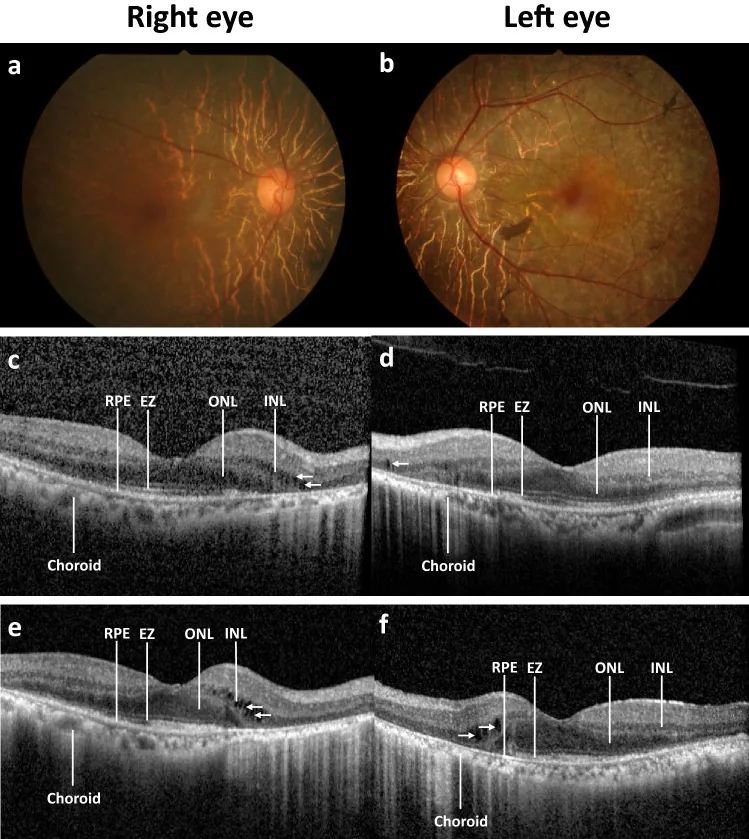
Disease progression of a patient with autoimmune retinopathy (AIR). Color fundus photography at baseline in the right (a) and left eye (b) shows normal optic disc, arteriolar attenuation, generalized retinal pigment epithelium (RPE) atrophy with macular sparing and scattered pigment clumps. Prominent large choroidal vessels can be observed around the optic disc. Optical coherence tomography images at baseline showing RPE attenuation, loss of ellipsoid zone (EZ) at the periphery, flattening of the outer nuclear layer (ONL), and slit cavitation in the inner nuclear layer (INL) on the nasal side, as marked by arrows, in the right (c) and left eye (d). The disease progression after five years is marked with RPE atrophy, progressive loss of EZ and ONL toward the fovea, more prominent slit cavit
Wang S, Hou H, Tang Y, Zhang S, Wang G, Guo Z, et al. An overview on CV2/CRMP5 antibody-associated paraneoplastic neurological syndromes. Neural regeneration research. 2023;18(11):2357-2364. doi:10.4103/1673-5374.371400. PMID:37282453; PMCID:PMC10360094.
Cross SA, Salomao DR, Parisi JE, Kryzer TJ, Bradley EA, Mines JA, et al. Paraneoplastic autoimmune optic neuritis with retinitis defined by CRMP-5-IgG. Annals of neurology. 2003;54(1):38-50. doi:10.1002/ana.10587. PMID:12838519.
Kaushik M, Virdee J, Giridharan S, Chavda SV, Batra R. Response of Recoverin-Positive Optic Neuritis to Chemotherapy, Steroid, and Plasma Exchange. Journal of neuro-ophthalmology : the official journal of the North American Neuro-Ophthalmology Society. 2024;44(1):e79-e81. doi:10.1097/WNO.0000000000001778. PMID:36729925.