Contact lens-induced dry eye (CLIDE) is a condition in which CL wear directly reduces tear film stability, causing ocular discomfort, visual disturbances, and corneal/conjunctival epithelial damage.
Dry eye is defined as “a chronic disease of the tears and ocular surface due to various factors, accompanied by ocular discomfort and visual disturbance” 7). CLIDE is a condition where CL wear is the primary cause among the “various factors” in this definition, and it involves a complex interplay of CL material, fitting, care solutions, and wearing time.
Many epidemiological studies have reported that CL use increases the risk of dry eye symptoms 7). On the other hand, the Osaka Study, based on dry eye diagnostic criteria, did not find CL wear to be a significant risk factor 7). While CLs can induce dry eye symptoms, evidence is inconsistent as to whether they are a risk factor for developing dry eye that meets diagnostic criteria.
Even with the widespread use of silicone hydrogel (SiHy) lenses today, approximately half of CL wearers are reported to have dry eye symptoms 7). The situation has changed with the evolution of materials, and the type of CLs used varies depending on the study period, which has a significant impact. TFOS DEWS III explicitly lists CL wear as a risk factor for dry eye and recommends changing CL material, design, replacement frequency, and care solutions for managing CLIDE 1).
The number of CL wearers worldwide is estimated to reach about 300 million 3), and the combined effect of digital devices and CL wear is a major risk for CLIDE, especially in young adults 4).
CLs affect all layers of the tear film: the lipid layer, aqueous layer, and mucin layer. Specifically, the following mechanisms are important.
CLs split the tear film into a pre-lens tear film (in front of the CL) and a post-lens tear film (behind the CL), making the pre-lens tear film thinner and more prone to evaporation.
The hydrophobic nature of CL materials (especially SiHy) leads to lipid deposits, destabilizing the tear lipid layer.
Mechanical friction during blinking damages the mucin layer.
Preservatives (e.g., benzalkonium chloride) cause goblet cell damage and reduce mucin production.
Chronic corneal nerve stimulation due to CL wear reduces reflex tear secretion.
QIs there a way to stop wearing CLs, such as alternatives to LASIK?
A
If dry eye symptoms become chronic due to CL wear, switching to refractive surgery (ICL, LASIK, SMILE) may be an option. ICL has the advantage of causing less postoperative dry eye because it does not ablate the cornea. However, suitability should be determined by an ophthalmologist, so it is advisable to first try to control symptoms and consult if that proves difficult.
Trobe J. Kellogg Eye Center, University of Michigan. Erosion of corneal epithelium owing to inadequate hydration. Figure 1. Wikimedia Commons, 2014. Source ID: Wikimedia Commons / File:Dry_eyeKell.jpg. License: CC BY 3.0.
Fluorescein staining observation of corneal epithelial erosion due to dehydration, showing epithelial defects stained fluorescent green. Corresponds to the corneal epithelial damage pattern by fluorescein staining discussed in the section “2. Main symptoms and clinical findings”.
The main subjective symptoms of CLIDE are as follows:
Subjective symptom
Characteristics/Frequency
Dryness/desert-like sensation
Worsens in the evening or after prolonged wear
Foreign body sensation/grittiness
Prominent in evaporative dry eye type
Redness
Mild to moderate, palpebral and bulbar conjunctiva
Blurred vision/visual fluctuation
Temporarily improves after blinking
Eye fatigue
Worsens after VDT work
Burning or stinging sensation
Worsened by eye drops or wind
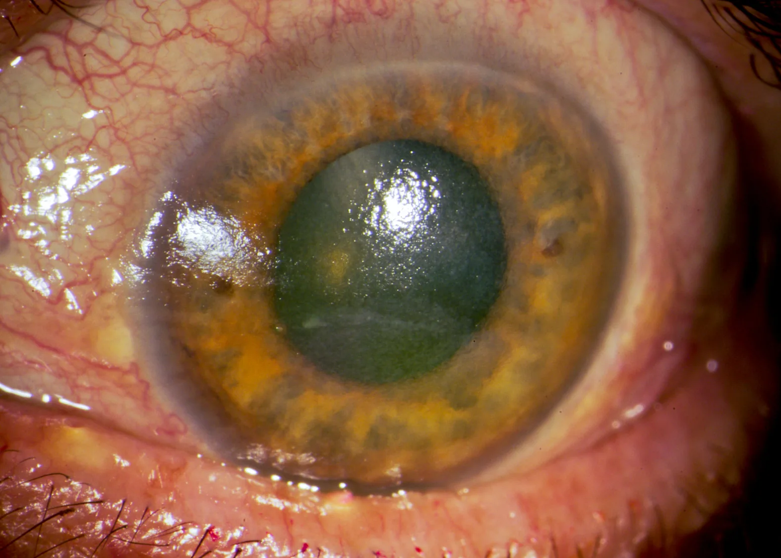
If all symptoms of redness, discharge, and pain are present, suspect infectious keratitis. Immediately remove the contact lens and seek medical attention.
Cause: Oxygen deficiency due to CL wear, or overall dryness from tear film instability
Course: Heals in 4–5 days with CL discontinuation and eye drops
Other findings include reduced tear meniscus (≤0.25 mm), shortened tear break-up time (BUT ≤5 seconds), and tear film instability due to lipid deposits in SiHy lens wearers.
Mechanical irritation and local dryness during blinking
HEMA-based SCL
Diffuse SPK and hypoxia
Corneal hypoxia due to low Dk/t
SiHy SCL
Smile mark SPK/SEAL
SEAL due to lipid deposits and hardness
Color CL
Donut-shaped SPK
Tear film instability at the pigmented area
Even after the widespread use of SiHy lenses, dry eye symptoms in wearers have not improved. The hydrophobicity of SiHy material makes it prone to lipid deposits, and the lipids adhering to the surface destabilize the lipid layer of the tear film. While conventional HEMA lenses mainly caused protein deposits, SiHy lenses produce deposits primarily composed of phospholipids and neutral lipids2).
TFOS DEWS III reports that lipid-containing artificial tears (lipomimetic eye drops) are effective in improving symptoms and corneal staining in CL wearers1).
During screen work, blink rate decreases markedly from about 16 times per minute to 5–7 times per minute, and incomplete blinks also increase4). Incomplete blinks prevent uniform spreading of the oil layer, leading to increased tear evaporation4). When VDT work and CL wear are combined, CLIDE is significantly exacerbated4).
VDT work is a risk factor for dry eye, with reports indicating that the risk of developing dry eye is 1.94 times higher in those who perform VDT work for more than 8 hours per day7). For CL wearers with long VDT work hours, it should be kept in mind that CLIDE is more likely to develop or worsen.
Preservatives such as benzalkonium chloride (BAK) can cause goblet cell damage and reduce the production of secretory mucins5). Mucin reduction leads to a tear film breakup pattern of poor wettability (spot/dimple break), which worsens CLIDE. Switching to preservative-free care products and eye drops can help reduce mucin damage.
Ring-shaped staining (which became a problem around 2010) has been reported with incompatibility between MPS (multipurpose solution) and SiHy lenses, so attention must be paid to the combination of care product type and lens material.
QDoes switching to daily disposable lenses improve CLIDE?
A
Daily disposable lenses require no lens care, have no risk of case contamination, and minimize accumulation of protein and lipid deposits. Care product-related complications and giant papillary conjunctivitis are less likely to occur. However, dry eye-related smile mark punctate keratopathy can still occur with daily disposable lenses, so it is important to also manage dry eye with eye drops and VDT work habits.
Based on the Japanese dry eye diagnostic criteria (2016 revision, Dry Eye Society). A definitive diagnosis of dry eye is made when both of the following two items are met7).
Subjective symptoms (ocular discomfort, visual function abnormalities) are present
Tear film breakup time (BUT) is 5 seconds or less
For the diagnosis of CLIDE, it is recommended to compare BUT while wearing CL and BUT after removing CL. If BUT is significantly shortened during CL wear, it is highly likely that CL is the trigger.
In CLIDE, evaporative type (random break) and aqueous-deficient type (spot/dimple break) are common. Pattern evaluation based on TFOD (Tear Film Oriented Diagnosis) directly guides treatment selection7).
The Dry Eye Clinical Practice Guideline recommends the use of artificial tears for dry eye in contact lens wearers 7). Depending on patient preference, changing lens material or lens care products, and oral omega-3 fatty acid supplementation are also suggested as treatment options 7) (strength of recommendation: weak, evidence level C).
TFOS DEWS III presents a stepwise treatment algorithm for evaporative dry eye disease including CLIDE 1). The algorithm escalates from lifestyle modification to eye drop therapy, then to punctal plugs and device therapy.
The following eye drops are currently covered by insurance in Japan and widely used.
Preservative-free artificial tears (e.g., Soft Santear): These form the foundation of treatment. Preservative-free formulations are preferred. Choose products that can be used while wearing contact lenses 7).
3% Diquafosol sodium ophthalmic solution (Diquas® / Diquas LX®) 6 times daily: A P2Y2 receptor agonist that promotes water and mucin secretion and oil layer spreading. Effective for both evaporative and aqueous-deficient dry eye. The Dry Eye Clinical Practice Guideline recommends its use (strong recommendation) 7). For use during contact lens wear, choose the preservative-free formulation (Diquas LX).
2% Rebamipide ophthalmic suspension UD (Mucosta® ophthalmic suspension UD) 4 times daily: Promotes mucin production and has anti-inflammatory effects. Particularly effective for aqueous-deficient dry eye (spot/dimple break). The Dry Eye Clinical Practice Guideline recommends its use (strong recommendation) 7). Single-use, preservative-free.
0.1% / 0.3% Sodium hyaluronate ophthalmic solution (Hyalein®) 6 times daily: Protects the corneal epithelium and retains moisture. During contact lens wear, avoid products containing preservatives 7).
Review of Contact Lens Materials and Care Products
If eye drops are insufficient, consider punctal plugs 7). However, in CLIDE where evaporative and aqueous-deficient types predominate, punctal plugs may cause tearing and blurred vision, so carefully assess indications.
If evaporative type predominates, warm compresses (5–10 minutes daily) and eyelid hygiene for MGD improve tear film stability 1). LipiFlow and IPL (intense pulsed light) are being studied for evaporative CLIDE with MGD background 1).
Oral omega-3 fatty acids have been reported to improve symptoms and BUT, but the evidence level is low and not covered by insurance, so they are an option as supplements 7).
QWhat should I do if dry eye symptoms persist even after removing contact lenses?
A
If symptoms persist after stopping CL use, dry eye may have become chronic triggered by CL wear. Follow dry eye treatment guidelines and continue secretagogues such as diquafosol sodium or rebamipide 7). For severe and refractory cases, consider punctal plugs, and add warm compresses if MGD is present. Consider resuming CL use only after dry eye is well controlled.
6. Pathophysiology and Detailed Mechanism of Onset
CLs affect all layers of the tear film: the lipid layer, aqueous layer, and mucin layer. The core pathophysiology of dry eye is the “vicious cycle of tear film instability and corneal/conjunctival epithelial damage,” and CLs accelerate this cycle.
When a CL is present, the tear film is divided into the pre-lens tear film (in front of the CL) and the post-lens tear film (behind the CL). The pre-lens tear film is thinner than normal and evaporates more easily. The post-lens tear film has reduced tear diffusivity due to contact with the hydrophobic surface of the CL.
SiHy materials are prone to lipid deposits due to their hydrophobicity, and the lipids adhering to the surface destabilize the lipid layer of the tear film. While conventional HEMA lenses mainly had protein deposits, SiHy lenses produce deposits primarily composed of phospholipids and neutral lipids 2). Tear film instability caused by lipid deposits is one reason why dry eye symptoms in CL wearers have not improved even after the widespread adoption of SiHy lenses.
During CL wear, incomplete blinks (blinks where the eyelids do not close completely) increase. In incomplete blinks, the lower cornea is not covered by tears for a period, which coincides with the location of smile-mark punctate keratopathy. When blink rate further decreases during VDT work (from typically 16 times/min to 5–7 times/min), CLIDE markedly worsens due to the synergistic effect with CL wear 4). In vivo confocal microscopy studies by Jalbert et al. have also shown corneal epithelial damage and reduced nerve fiber density due to CL wear 14), suggesting that chronic CL wear affects corneal nerves, leading to decreased tear secretion and worsening of CLIDE.
Goblet Cell Damage by Preservatives in Care Solutions
Preservatives such as benzalkonium chloride (BAK) cause goblet cell damage and reduce the production of secretory mucin 5). Mucin deficiency leads to a wetting-deficiency type of tear breakup pattern (spot/dimple break), which worsens CLIDE. Switching to preservative-free care solutions and eye drops can help reduce mucin damage.
A vicious cycle is formed: tear film instability → epithelial damage → inflammatory cytokine production → goblet cell damage → mucin deficiency → tear film instability. CLs directly trigger the entry point of this cycle (tear film destabilization). Chronic stimulation of corneal sensory nerves leads to reduced sensation, further decreasing reflex tear secretion and accelerating the cycle.
Meibomian gland dysfunction (MGD) is a major exacerbating factor for CLIDE. When MGD is present, the tear lipid layer becomes thinner, leading to pronounced evaporative CLIDE. Daily warm compresses (5–10 minutes, approximately 42°C) and eyelid hygiene can improve MGD, stabilize the oil layer, and alleviate CLIDE symptoms 1).
Warm compresses and eyelid hygiene as treatment for MGD are the most basic and effective interventions to break the “vicious cycle” of CLIDE. In particular, for SiHy lens wearers with evaporative tear instability, it is desirable to prioritize MGD treatment before changing the CL material. For eyelid hygiene, use a dedicated eyelid cleanser (eyelid-specific cleaning product) and gently clean the meibomian gland openings 1–2 times a day.
Long-term CL wear reduces corneal sensation (hypoesthesia). Due to reduced sensation, CL wearers may not notice early symptoms of dry eye, and epithelial damage can progress unnoticed. As hypoesthesia advances, reflex tear secretion further decreases, leading to chronic CLIDE. Hypoesthesia is particularly pronounced in HCL wearers, but also occurs in SCL wearers with long-term use. To prevent chronic CLIDE, regular slit-lamp examination and tear film testing are important regardless of symptoms.
With appropriate CL material change, improved care, and eye drop treatment, most cases of CLIDE can be symptom-controlled. However, once CLIDE is established, background factors such as corneal hypoesthesia, MGD, and decreased goblet cell density may persist even after CL discontinuation, so careful evaluation is necessary before resuming CL wear.
Eye drop treatment is more likely to show symptom improvement when continued for at least 4 weeks, and improvement in findings (e.g., corneal staining) may require 2–4 months of continuous use 1). It is important to explain to patients that “effects may not appear immediately” to maintain adherence.
In managing CLIDE, controlling wearing time is a non-pharmacological intervention as important as drug therapy.
Guideline for Wearing Time
Recommendations
Daily wearing time
Generally within 8–10 hours
Removal time before bedtime
Remove CL 2–3 hours before bedtime to allow tear film recovery
Wear during sleep
Absolutely prohibited (exacerbation of CLIDE + infection risk)
Regular check-ups
Ophthalmology visits 1–2 times per year are mandatory
For patients who need to wear contact lenses for long periods (e.g., long-time VDT workers), it is effective to instruct them to take “hydration breaks” by removing the lenses and switching to glasses midway. Also recommend carrying preservative-free eye drops (e.g., Diquas LX, Hyalein preservative-free) for use.
TFOS DEWS III (2025) identifies CL wear as a risk factor for dry eye and recommends changes in CL material, design, replacement frequency, and care solutions for the management of CLIDE 1). It also reports that lipid-containing artificial tears (lipomimetic eye drops) are effective in improving symptoms and corneal staining in CL wearers 1).
The TFOS Lifestyle Report states that there are approximately 300 million CL wearers worldwide, and that wearing CLs destabilizes the tear film and increases the risk of DED symptoms 3). Steele et al.’s review updates the epidemiology of CL-related infiltrative events, clearly positioning CLIDE as a risk factor for CIE 15). The combined effect of digital devices and CL wear is a major risk for CLIDE onset, especially in young adults 4).
With the widespread use of multifocal soft CLs and orthokeratology for myopia progression control, managing CLIDE in children and adolescents will become an important issue. Although Level I RCT evidence for multifocal CLs is accumulating, the evaluation of dry eye risk associated with long-term wear is still insufficient 6).
LipiFlow (vectored thermal pulsation therapy) and IPL (intense pulsed light) are being studied as treatment options for evaporative CLIDE associated with MGD1). Since evaporation is a key mechanism of CLIDE, the combination of MGD treatment and CL fitting is expected to gain attention.
It has been suggested that ocular surface weakening and reduced tear protection due to CLIDE may predispose to Acanthamoeba keratitis (AK). Switching to DD lenses eliminates poor care and is reported to reduce AK risk by approximately 3.84 times compared to DW reusable lenses 8). In managing CLIDE, changing to DD lenses is also an effective strategy for preventing care solution-related complications.
Association with CL-Related Non-Infectious Corneal Infiltrates
When CLIDE becomes severe, the risk of CL-related non-infectious corneal infiltrative events (CIE) increases. CIE includes asymptomatic infiltrates (AI), CLARE, etc. 11), with an annual incidence of approximately 3–6 per 100 person-years 12). With EW of SiHy lenses, the annual CIE incidence reaches about 20 per 100 person-years 12), and CLIDE and CIE should be understood as a continuous disease spectrum.
Lens case contamination is also one of the exacerbating factors of CLIDE. Bacterial contamination is found in 30–80% of lens cases in use 13), and endotoxins derived from contaminating bacteria may be continuously exposed to the ocular surface, potentially worsening CLIDE.
In patients suspected of having CLIDE, according to the Infectious Keratitis Clinical Practice Guidelines (3rd edition), if the triad of hyperemia, discharge, and pain is present, infectious keratitis should be considered first, and corneal culture testing should be performed 9).
According to the definition of the TFOS International Workshop on Contact Lens Discomfort, contact lens discomfort is defined as “discomfort that in principle continuously worsens with contact lens wear” 10), and CLIDE is positioned as a major causative disease of this CL discomfort. In patients complaining of persistent CL discomfort, actively evaluating and treating CLIDE is important for improving the rate of continued lens wear.
Jones L, Craig JP, Markoulli M, Karpecki P, Akpek EK, Basu S, et al. TFOS DEWS III: Management and Therapy. American journal of ophthalmology. 2025;279:289-386. doi:10.1016/j.ajo.2025.05.039. PMID:40467022.
Stapleton F, Bakkar M, Carnt N, Chalmers R, Vijay AK, Marasini S, et al. CLEAR - Contact lens complications. Contact lens & anterior eye : the journal of the British Contact Lens Association. 2021;44(2):330-367. doi:10.1016/j.clae.2021.02.010. PMID:33775382.
Craig JP, Alves M, Wolffsohn JS, Downie LE, Efron N, Galor A, et al. TFOS Lifestyle Report Executive Summary: A Lifestyle Epidemic - Ocular Surface Disease. The ocular surface. 2023;30:240-253. doi:10.1016/j.jtos.2023.08.009. PMID:37659474.
Wolffsohn JS, Lingham G, Downie LE, et al. TFOS Lifestyle: Impact of the digital environment on the ocular surface. Ocul Surf. 2023;28:213-252. doi:10.1016/j.jtos.2023.04.004.
American Academy of Ophthalmology. Bacterial Keratitis Preferred Practice Pattern. Ophthalmology. 2024;131(2):P265-P330.
Cavuoto KM, Trivedi RH, Prakalapakorn SG, Oatts JT, Nallasamy S, Morrison DG, Pineles SL, Chang MY. Multifocal Soft Contact Lenses for the Treatment of Myopia Progression in Children: A Report by the American Academy of Ophthalmology. Ophthalmology. 2025;132(4):495-503. doi:10.1016/j.ophtha.2024.09.031. PMID:39503665; PMCID:PMC11930616.
Dumbleton K, Caffery B, Dogru M, Hickson-Curran S, Kern J, Kojima T, et al. The TFOS International Workshop on Contact Lens Discomfort: report of the subcommittee on epidemiology. Investigative ophthalmology & visual science. 2013;54(11):TFOS20-36. doi:10.1167/iovs.13-13125. PMID:24058130.
Sweeney DF, Jalbert I, Covey M, Sankaridurg PR, Vajdic C, Holden BA, et al. Clinical characterization of corneal infiltrative events observed with soft contact lens wear. Cornea. 2003;22(5):435-42. doi:10.1097/00003226-200307000-00009. PMID:12827049.
Loretta Szczotka‐Flynn, Mireya Diaz. Risk of Corneal Inflammatory Events with Silicone Hydrogel and Low Dk Hydrogel Extended Contact Lens Wear: A Meta‐Analysis. OVS. 2007;84(4):247-256. doi:10.1097/opx.0b013e3180421c47.
Wu YT, Willcox M, Zhu H, Stapleton F. Contact lens hygiene compliance and lens case contamination: A review. Contact lens & anterior eye : the journal of the British Contact Lens Association. 2015;38(5):307-16. doi:10.1016/j.clae.2015.04.007. PMID:25980811.
Jalbert I, Willcox MD, Sweeney DF. Isolation of Staphylococcus aureus from a contact lens at the time of a contact lens-induced peripheral ulcer: case report. Cornea. 2000;19(1):116-20. doi:10.1097/00003226-200001000-00023. PMID:10632021.
Steele KR, Szczotka-Flynn L. Epidemiology of contact lens-induced infiltrates: an updated review. Clinical & experimental optometry. 2017;100(5):473-481. doi:10.1111/cxo.12598. PMID:28868803.
Copy the article text and paste it into your preferred AI assistant.
Article copied to clipboard
Open an AI assistant below and paste the copied text into the chat box.