Dry eye is defined as “a disease in which the stability of the tear film is reduced due to various factors, causing ocular discomfort and visual dysfunction, and may be accompanied by ocular surface damage.” Dry eye after refractive surgery is primarily caused by corneal nerve transection and disruption of the lacrimal gland reflex arc due to corneal surgery, among the “various factors” in this definition.
It has been suggested that corneal epithelial damage and decreased tear film breakup time (BUT) are likely to be induced by anterior segment surgeries in general, including LASIK and PRK. Tear secretion (Schirmer value) has been suggested to decrease in surgeries involving corneal incision or excision 2).
In the guidelines for refractive surgery (8th edition), dry eye is clearly listed as one of the complications after LASIK 1), and preoperative evaluation and active management are required.
Postoperative dry eye symptoms generally improve within 6 to 12 months, but in a small number of cases, they progress to refractory dry eye that does not respond to conventional treatment 3).
QWhy is dry eye more likely after LASIK?
A
In LASIK, when creating a corneal flap with a microkeratome or femtosecond laser, the corneal nerve plexus in the anterior stroma is extensively severed. This significantly blocks reflex signals to the lacrimal gland via the trigeminal nerve, reducing both basal and reflex secretion. Additionally, decreased blinking and worsening of meibomian gland dysfunction after surgery contribute to many patients experiencing dry eye symptoms for 1 to 3 months postoperatively.
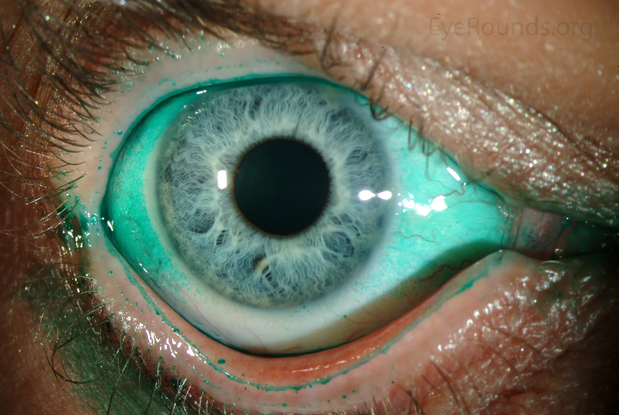
Vislisel J, Critser B. Diffuse lissamine green staining in severe keratoconjunctivitis sicca. EyeRounds.org Atlas, University of Iowa, 2016. Source ID: Wikimedia Commons / File:LG3-LRG.jpg. License: CC BY-SA 4.0.
Ocular surface of severe dry eye observed with lissamine green staining, showing diffuse epithelial staining over the entire conjunctiva and punctate staining of the corneal epithelium. This corresponds to the corneal and conjunctival epithelial damage findings by lissamine green and fluorescein staining discussed in the section “Main symptoms and clinical findings.”
The main mechanisms of dry eye after LASIK are as follows:
During flap creation, the corneal nerve plexus (derived from the ophthalmic branch of the trigeminal nerve) in the anterior stroma is extensively severed
Reflex signals to the lacrimal gland via the trigeminal nerve are interrupted → decreased basal and reflex secretion
Decreased tear volume and stability → vicious cycle of corneal and conjunctival epithelial damage
Temporary worsening of meibomian gland function due to postoperative inflammation → adds an evaporative component
Risk Comparison by Surgical Procedure
LASIK (flap method): Extensive transection of the anterior stromal nerve plexus during flap creation. Highest incidence of postoperative dry eye. Incidence 50–90% (short-term postoperative)
PRK (surface ablation): The subepithelium is ablated, but no flap is created, so corneal nerve preservation is higher than LASIK. Early postoperative BUT shortening occurs due to epithelial debridement, but long-term effects are milder than LASIK
SMILE (small incision lenticule extraction): Small incision (2–3 mm) minimizes corneal nerve transection. Postoperative dry eye is reported to be milder than LASIK. Nerve recovery after SMILE has been reported to be faster than after FS-LASIK4)
Preoperative Risk Factors
Preoperative dry eye: Patients with dry eye before surgery are suggested to have more severe dry eye after LASIK compared to those without dry eye2)
Female and elderly: General risk factors for dry eye also affect the postoperative period.
High myopia correction (large ablation depth): Deeper ablation reduces the preservation rate of corneal nerves.
After refractive surgery, regular preoperative and postoperative evaluation of BUT and subjective symptoms allows objective assessment of postoperative changes.
Corneal sensitivity test is a specific test for dry eye after refractive surgery and is useful for evaluating the degree of corneal nerve severance after surgery.
Subtype diagnosis by TFOD (Tear Film Oriented Diagnosis)
Observe the tear film breakup pattern immediately after eye opening to identify the dry eye subtype. In postoperative dry eye, a mixture of spot/dimple break (poor wettability type) and line break (tear deficiency type) is common, enabling a diagnosis directly linked to TFOT.
Before refractive surgery, it is recommended to evaluate the following and initiate dry eye treatment preoperatively 3).
Subjective symptom scoring such as OSDI (OSDI ≥13 indicates mild dry eye)
BUT and Schirmer test (BUT ≤5 seconds or Schirmer ≤5 mm/5 min indicates need for preoperative treatment)
Meibography and eyelid margin evaluation (to check for MGD and its severity)
Corneal and conjunctival staining (lissamine green, fluorescein)
Corneal sensitivity test (Cochet-Bonnet)
Non-invasive tear break-up time (NIBUT)
Detecting and actively treating dry eye preoperatively improves postoperative visual outcomes and patient satisfaction 3). In particular, when MGD is present, preoperative IPL treatment, LipiFlow, or warm compresses have been reported to significantly improve OSDI and BUT at 3 months postoperatively 3). Preoperative ocular surface optimization is one of the most effective strategies for preventing postoperative dry eye. The Refractive Surgery Guidelines (8th edition) 1) list dry eye as a factor requiring caution, and dry eye syndrome is an absolute contraindication for SMILE.
QIf I want to undergo refractive surgery but have dry eye, can I still have the surgery?
A
Even if dry eye is present preoperatively, the efficacy and safety of LASIK are considered comparable to those in patients without dry eye2). However, patients with preoperative dry eye tend to have more severe dry eye after LASIK, so it is important to actively treat dry eye before surgery and optimize the ocular surface. In cases of MGD (meibomian gland dysfunction), preoperative treatment with LipiFlow or warm compresses has been reported to significantly improve OSDI and BUT at 3 months postoperatively 3).
Note the specificity of postoperative dry eye: it may be complicated by “neurotrophic dry eye” due to decreased corneal sensation (nerve transection). In cases with decreased sensation, in addition to standard TFOT treatment, it is effective to add adjunctive treatments that promote nerve recovery (e.g., omega-3 fatty acids, PRGF, autologous serum eye drops) 3).
First-line treatment for postoperative dry eye is preservative-free artificial tears and mucin secretagogues.
3% Diquafosol sodium ophthalmic solution (Diquas®):
P2Y2 receptor agonist. It has a combined effect of promoting water and mucin secretion and oil layer spreading. Its efficacy for postoperative dry eye has been demonstrated, and the Dry Eye Clinical Practice Guideline gives a “recommend to perform” (strong recommendation) 2). In an RCT of 61 eyes undergoing femtosecond LASIK, 3% diquafosol + 0.15% HA eye drops significantly improved OSDI, TBUT, and lipid layer compared to 0.15% HA alone 3).
2% Rebamipide ophthalmic suspension UD (Mucosta® ophthalmic suspension UD):
Promotes mucin production and has anti-inflammatory effects. In an RCT of 60 eyes with dry eye after corneal refractive surgery, rebamipide 4 times/day significantly improved Schirmer value, BUT, corneal staining, and irregularity index compared to artificial tears 3).
0.1% / 0.3% Sodium hyaluronate ophthalmic solution (Hyalein®):
Corneal epithelial protection and water retention. Preservative-free formulations (Hyalein Mini®) are preferable. TFOS DEWS III confirmed that many tear supplements including HA improve symptoms and signs of postoperative dry eye5).
Prescription Example (Mild to Moderate Postoperative)
Indicated for aqueous-deficient dry eye that does not respond adequately to eye drops. The Dry Eye Clinical Practice Guideline recommends punctal plugs as “perform”, and improvement in symptoms has been reported before and after punctal plug treatment for post-LASIK dry eye2).
Silicone plugs: Super Eagle™ plug, Punctal Plug® F, etc. Provide permanent occlusion.
Liquid collagen plug (Keeptear®): A liquid plug made of atelocollagen. It gels at body temperature (36°C) and occludes the canaliculus. Suitable for temporary exacerbation of dry eye. Store refrigerated at 2–10°C.
If MGD is complicated with postoperative dry eye, it should be actively treated3).
Warm compress and eyelid hygiene: Eyelid massage after a hot towel (40–42°C, 5–10 minutes). Basic care for MGD.
IPL (intense pulsed light): Two prospective studies evaluated its efficacy in LASIK-induced refractory dry eye (moderate to severe cases unresponsive to conventional treatment for over 1 year). One study (42 eyes) showed significant improvement in NIBUT, OSDI, tear lipid layer, and meibomian gland function after two IPL sessions. Another RCT (50 cases) found that IPL combined with a warm eye mask was more effective than IPL alone in improving subjective and objective parameters3).
Effect of preoperative MGD treatment: In patients with pre-existing MGD before LASIK (32 cases), vector thermal pulsation therapy performed 1 week before LASIK significantly improved OSDI and BUT at 3 months postoperatively3).
In a masked RCT of 61 patients who received three IPL sessions preoperatively (before surgery, 1 week after, and 3 weeks after), the IPL group showed significant improvement in OSDI, NIBUT, TMH, and meibography at 3 months postoperatively, while the control group had decreased TMH. At 6 months, the difference in OSDI narrowed, but objective tear parameters remained improved3).
IPL delivers light pulses of 532–1200 nm to the periorbital area, occluding capillaries to reduce chronic inflammation around the meibomian glands and improve gland secretion. Typically, 4–5 sessions (3–4 weeks apart) constitute one course of treatment. The effect is maximized by the physician expressing the meibomian glands after treatment. Pigmented skin lesions, Fitzpatrick skin type VI, and eyes that have undergone refractive surgery within the past 4 weeks are contraindications or require caution for IPL.
QWhat treatments are effective for refractory dry eye after LASIK?
A
For refractory dry eye after LASIK that does not respond to conventional eye drops, IPL (intense pulsed light), warm compresses, and vector thermal pulsation therapy (LipiFlow) are considered effective3). IPL improves meibomian gland function, NIBUT, OSDI, and the lipid layer. Combining IPL with a warm eye mask is more effective than IPL alone. Autologous serum eye drops (PRGF) are also an option for refractory cases.
The sensory nerves of the cornea are mainly composed of the corneal nerve plexus derived from the nasociliary nerve, a branch of the ophthalmic division of the trigeminal nerve (V1). These nerves form a dense plexus in the anterior stroma and provide the main sensory input to the cornea.
During LASIK, flap creation (using a microkeratome or femtosecond laser) cuts the anterior stromal nerve plexus extensively in a circumferential direction. This nerve transection triggers the following cascade.
Reflex arc disruption:
Corneal surface stimulation (afferent pathway) → trigeminal nucleus → parasympathetic innervation to the lacrimal gland (efferent pathway) is interrupted
Reflex tear secretion (both basal and reflex) is significantly reduced
Decreased tear volume → drying of the corneal and conjunctival epithelium → shortened tear break-up time (BUT)
Effect on membrane-bound mucins:
Corneal nerves also supply neurotrophic factors to the corneal epithelium. After nerve transection, the supply of neurotrophic factors (such as EGF and NGF) decreases, leading to reduced expression of membrane-bound mucin (MUC16). Decreased mucin expression → reduced wettability → spot/dimple break (wettability-deficient dry eye) occurs.
Course of nerve regeneration:
Corneal nerves gradually regenerate over 6 to 12 months after surgery
After LASIK, regeneration takes time due to 360° nerve transection. In many cases, about 80% recovery occurs within one year, but some cases may take several years
After SMILE, nerve density recovery is faster because only a small 2–3 mm incision is made4)
In vivo confocal microscopy (IVCM) allows objective monitoring of nerve recovery by evaluating corneal nerve density over time
If corneal hypoesthesia persists for more than 6 months, there is a risk of progression to neurotrophic keratopathy, and the indication for nerve growth factor (NGF) eye drops should be considered
SMILE uses only a 2–3 mm small incision to extract the intrastromal lenticule, minimizing circumferential nerve damage. Comparative studies of FS-LASIK and SMILE have reported that SMILE leads to faster recovery of corneal nerve density and less impact on tear parameters than FS-LASIK4).
Surgery-related inflammation temporarily worsens MGD function and promotes evaporative dry eye. Infiltration of inflammatory cytokines (MMP-9, IL-1β, etc.) into the conjunctiva and corneal stroma after LASIK contributes to tear film instability. Cyclosporine eye drops have been reported to significantly improve OSDI, BUT, and corneal sensitivity after various refractive surgeries including LASIK3).
Postoperative inflammation usually resolves spontaneously within 1–3 months, but in patients with preoperative allergic conjunctival disease, MGD, or steroid responders, inflammation may persist or worsen. If long-term steroid eye drops are needed after surgery, regular intraocular pressure monitoring is necessary, and attention should be paid to possible association with IFS (interface fluid syndrome).
Relationship Between Corneal Shape Changes and Ocular Surface
Changes in corneal shape after refractive surgery can affect the contact pattern between the upper eyelid and the cornea (lid-globe apposition), altering tear distribution and spreading. In particular, corneal flattening after high myopia correction may impair tear spreading.
Additionally, postoperative corneal hypoesthesia reduces the blink reflex and decreases the number of complete blinks. This impairs mechanical tear spreading and oil layer redistribution, promoting evaporative dry eye. Complete blink training (conscious practice of full blinking) is recommended as a self-care measure for postoperative dry eye management3).
TFOS DEWS III (2025) recommends optimization of the ocular surface before and after corneal refractive surgery as standard care5). Several prospective studies have shown that preoperative intervention in patients with evaporative dry eye (MGD) improves postoperative ocular surface status and visual parameters3).
TFOS DEWS III also summarizes the latest evidence on next-generation tear supplements (e.g., perfluorohexyloctane, silk-derived proteins, rhPRG4), which are expected to become additional treatment options alongside standard HA eye drops5).
There are reports that botulinum toxin injection into the medial lower eyelid improves DED symptoms and signs. Furthermore, a study showed that botulinum toxin significantly improved dry eye symptoms after LASIK with fewer complications than punctal plugs or eye drops, and it is attracting attention as a future option 3). The mechanism is thought to be that botulinum toxin suppresses contraction of the lacrimal canaliculi, delaying tear drainage and increasing the volume of tears retained on the ocular surface. However, there are risks of side effects such as ptosis and diplopia, and administration requires a specialized facility.
In a retrospective comparative study (77 eyes) of dry eye patients after LASIK, PRGF eye drops significantly improved visual acuity, TBUT, OSDI, symptom scores, and Schirmer values compared to conventional artificial tears 3). PRGF contains growth factors (EGF, NGF, etc.) and may promote corneal nerve recovery. Autologous serum eye drops are similar biological eye drops prepared from the patient’s own serum and have shown efficacy for refractory dry eye associated with corneal neuropathy. However, preparation and use of autologous serum and PRGF eye drops require protocol management at a specialized facility 3).
An association between omega-3 fatty acid intake and improvement in corneal nerve parameters has been suggested, and it is attracting attention as an aid for corneal nerve recovery after refractive surgery 5). Research on neurotrophic factor (NGF) eye drops for neurotrophic keratopathy and postoperative corneal neuropathy is also ongoing.
TFOS DEWS III has indicated that low-level light therapy (LLLT) may have similar effects to IPL for dry eye with MGD, and research is progressing as a new non-contact, painless treatment option 5). LLLT irradiates the periorbital area with near-infrared light of 633–850 nm, improving meibomian gland function and having anti-inflammatory and tissue regeneration-promoting effects. Clinical evidence for its application to postoperative dry eye is still limited, but it is expected as an option for patients for whom IPL is contraindicated (e.g., those with pigmented lesions) or who desire a less invasive treatment 5).
Postoperative Dry Eye Management in KLEx Guidelines
In the KLEx (small incision lenticule extraction) international guidelines by Wang et al. 6), evidence-based recommendations for ocular surface management after SMILE are provided, including preoperative dry eye evaluation and active postoperative eye drop treatment. Although SMILE has a lower risk of postoperative dry eye than LASIK, it is considered best to improve the preoperative dry eye state before performing surgery 6).
Association Between Corneal Ectasia PPP and Dry Eye
The AAO Corneal Ectasia PPP7) recognizes the complication of dry eye associated with corneal nerve damage in the management of postoperative ectasia, and parallel management of irregular astigmatism correction (RGP, scleral lenses, etc.) and dry eye treatment is necessary. Scleral lenses are positioned as an effective option for both correcting irregular astigmatism and improving dry eye symptoms7).
The mathematical model by Reinstein et al.8) showed that SMILE may relatively preserve the anterior corneal stroma. The association with postoperative dry eye needs to be interpreted not only from this mechanical model but also in combination with clinical studies dealing with corneal nerves and tear film indicators4, 8).
The systematic review by Moshirfar et al.9) organized the risk assessment of postoperative ectasia. In surgical technique selection, ectasia risk assessment and preoperative dry eye evaluation should be performed separately, and the decision should be made comprehensively based on corneal shape, ablation amount, and tear film status.
Combined management of progressive corneal ectasia and dry eye
The international consensus by Gomes et al.10) indicates that patients with corneal ectasia have a high rate of dry eye comorbidity, and it is recommended to actively perform tear supplementation and anti-inflammatory treatment in ocular surface management before and after CXL (corneal cross-linking)10).
Igarashi et al.11) reported in an RCT targeting dry eye after corneal refractive surgery that 2% rebamipide ophthalmic solution significantly improved Schirmer value, BUT, corneal staining score, and irregular astigmatism index compared to artificial tears. Rebamipide has a combined action of promoting mucin production and anti-inflammation, and has been shown to be effective for both aqueous-deficient and mucin-deficient types of postoperative dry eye11).
Implications for ocular surface management from the KERALINK trial
The KERALINK trial by Larkin et al.12) demonstrated the efficacy of CXL in young keratoconus patients, and continuous use of tear supplements was recommended during the trial, reaffirming the importance of combined management of corneal ectasia and dry eye12).
Santhiago et al.13) showed that PTA ≥ 40% is a risk factor for postoperative ectasia. Since deep ablation can also affect corneal nerves, it is reasonable to evaluate ectasia risk and dry eye risk in parallel when designing the surgical procedure4, 13).
Integration of Randleman risk score and preoperative dry eye evaluation
The ectasia risk scoring system by Randleman et al.14) is a framework that evaluates corneal shape, residual stromal thickness, and degree of myopia. Preoperatively, dry eye is assessed separately using BUT, Schirmer test, and OSDI to comprehensively estimate the risk of postoperative complications.
Association between forme fruste corneal ectasia and dry eye
Ectasia after LASIK in forme fruste keratoconus reported by Seiler et al.15) is an early report indicating the importance of preoperative evaluation of latent corneal ectasia. The risk of severe dry eye is assessed preoperatively separately from corneal shape risk.
Jones L, Craig JP, Markoulli M, Karpecki P, Akpek EK, Basu S, et al. TFOS DEWS III: Management and Therapy. American journal of ophthalmology. 2025;279:289-386. doi:10.1016/j.ajo.2025.05.039. PMID:40467022.
Recchioni A, Sisó-Fuertes I, Hartwig A, Hamid A, Shortt AJ, Morris R, et al. Short-Term Impact of FS-LASIK and SMILE on Dry Eye Metrics and Corneal Nerve Morphology. Cornea. 2020;39(7):851-857. doi:10.1097/ICO.0000000000002312. PMID:32243424.
Jones L, Craig JP, Markoulli M, Karpecki P, Akpek EK, Basu S, Bitton E, Chen W, et al. TFOS DEWS III: Management and Therapy. American journal of ophthalmology. 2025;279:289-386. doi:10.1016/j.ajo.2025.05.039. PMID:40467022.
Wang Y, Xie L, Yao K, Sekundo W, Alió JL, Mehta JS, Goel S, Elmassry A, Schallhorn J, Shilova T, Cao H, Xu L, Chen X, Zhang F, Bai J, Zhang W, Liu Q, Zhou X, Chen Y, Wang Z, Jhanji V, Yang K, Writing Committee for the Guideline Working Group. Evidence-Based Guidelines for Keratorefractive Lenticule Extraction Surgery. Ophthalmology. 2025;132(4):397-419. doi:10.1016/j.ophtha.2024.11.016. PMID:39577672.
Jhanji V, Ahmad S, Amescua G, et al. Corneal Ectasia Preferred Practice Pattern. Ophthalmology. 2024 Apr;131(4):P205-P246. doi:10.1016/j.ophtha.2023.12.038. PMID:38349299.
Reinstein DZ, Archer TJ, Randleman JB. Mathematical model to compare the relative tensile strength of the cornea after PRK, LASIK, and small incision lenticule extraction. J Refract Surg. 2013;29:454-460. doi:10.3928/1081597X-20130617-03. PMID:23820227.
Moshirfar M, Tukan AN, Bundogji N, Liu HY, McCabe SE, Ronquillo YC, et al. Ectasia After Corneal Refractive Surgery: A Systematic Review. Ophthalmology and therapy. 2021;10(4):753-776. doi:10.1007/s40123-021-00383-w. PMID:34417707; PMCID:PMC8589911.
Gomes JA, Tan D, Rapuano CJ, Belin MW, Ambrósio R, Guell JL, Malecaze F, Nishida K, Sangwan VS, Group of Panelists for the Global Delphi Panel of Keratoconus and Ectatic Diseases. Global consensus on keratoconus and ectatic diseases. Cornea. 2015;34(4):359-369. doi:10.1097/ico.0000000000000408. PMID:25738235.
Igarashi T, Kamiya K, Kobashi H, Shimizu K. Effect of Rebamipide Ophthalmic Suspension on Intraocular Light Scattering for Dry Eye After Corneal Refractive Surgery. Cornea. 2015;34(8):895-900. doi:10.1097/ICO.0000000000000456.
Larkin DFP, Chowdhury K, Burr JM, et al. Effect of corneal cross-linking versus standard care on keratoconus progression in young patients: The Keralink randomized controlled trial. Ophthalmology. 2021;128:1516-1526. doi:10.1016/j.ophtha.2021.04.019. PMID:33892046.
Santhiago MR, Smadja D, Gomes BF, et al. Association between the percent tissue altered and post-LASIK ectasia in eyes with normal preoperative topography. Am J Ophthalmol. 2014;158:87-95.e1. doi:10.1016/j.ajo.2014.04.002. PMID:24727263.
Randleman JB, Woodward M, Lynn MJ, Stulting RD. Risk assessment for ectasia after corneal refractive surgery. Ophthalmology. 2008 Jan;115(1):37-50.e4. doi:10.1016/j.ophtha.2007.03.073. PMID:17624434.
Theo Seiler, Andreas W. Quurke. Iatrogenic keratectasia after LASIK in a case of forme fruste keratoconus. Journal of Cataract and Refractive Surgery. 1998;24(7):1007-1009. doi:10.1016/s0886-3350(98)80057-6.
Copy the article text and paste it into your preferred AI assistant.
Article copied to clipboard
Open an AI assistant below and paste the copied text into the chat box.