
Grade I (pre-steal)
Decreased vertebral artery blood flow: Antegrade blood flow in the affected vertebral artery is reduced.
No subjective symptoms: Often discovered incidentally on imaging or ultrasound.

Subclavian steal syndrome (SSS) is a condition in which stenosis or occlusion of the proximal subclavian artery causes reversal of blood flow in the ipsilateral vertebral artery. This leads to hypoperfusion of the vertebrobasilar system, reducing blood flow to the occipital lobe, brainstem, and eyes, resulting in various neurological and ocular symptoms.
In 1960, Contorni reported the first case of angiographic reflux in a patient with absent radial pulse, and in 1961, Fisher coined the term “subclavian steal”.
Epidemiology
It is found in 2–4% of the general population, but most cases are asymptomatic3). Among patients with peripheral arterial disease, about 30% have subclavian artery stenosis, and even with a large blood pressure difference, many remain asymptomatic4).
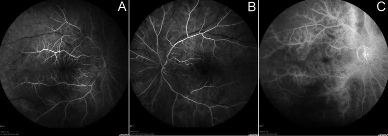
Subjective symptoms of SSS reflect hypoperfusion in the vertebrobasilar artery system.
Systemic Findings
Ophthalmic Findings
Severity Classification
Grade I (pre-steal)
Decreased vertebral artery blood flow: Antegrade blood flow in the affected vertebral artery is reduced.
No subjective symptoms: Often discovered incidentally on imaging or ultrasound.
Grade II (alternating flow)
Diastolic antegrade, systolic retrograde: The direction of blood flow changes with the pulse.
Mild to moderate symptoms: Symptoms tend to appear during exercise.
Grade III (Persistent Reversal)
Constant retrograde flow: Blood flow in the affected vertebral artery is continuously reversed.
Prominent symptoms: Hypoperfusion symptoms in the vertebrobasilar system occur even at rest.
A systolic blood pressure difference of 20 mmHg or more raises suspicion of SSS. A difference of 40 mmHg or more corresponds to Grade II–III, but there are cases with a 70 mmHg difference that remain asymptomatic 4). It is difficult to predict symptoms based solely on blood pressure difference, and confirmation by ultrasound or imaging is necessary.
The most common cause of SSS is atherosclerosis, and 81% of patients are reported to have multiple atherosclerotic lesions in the cerebral supply vessels.
Other causes
Risk factors
Special conditions
Excessive blood flow from dialysis access (such as a shunt) can cause SSS-like symptoms even without subclavian artery stenosis 1). Dizziness and upper limb symptoms during dialysis may be due to this mechanism and require early evaluation.
The characteristics of each imaging diagnostic method are shown below.
| Examination method | Main use | Features |
|---|---|---|
| CTA | Identification of stenosis and calcification assessment | Visualizes occlusion and retrograde filling3) |
| MRA (TOF method) | Vertebral artery signal evaluation | Decreased signal on the affected side is useful for early diagnosis5) |
| DSA (cerebral angiography) | Definitive diagnosis and treatment | Gold standard6) |
Tanaka et al. (2022) reported a case of a 76-year-old male with recurrent vertigo, in which TOF-MRA showed decreased signal in the left proximal intracranial vertebral artery, and angiography confirmed occlusion of the left subclavian artery origin and left vertebral artery reflux 5). They reported that changes in MRA signal intensity are useful for early diagnosis of SSS.
Management of risk factors and pharmacotherapy are the mainstays.
Ophthalmic treatment for ocular ischemia
The current first-line treatment is percutaneous transluminal angioplasty (PTA) with stent placement.
Neupane et al. (2024) performed angioplasty plus stent placement for severe proximal left subclavian artery occlusion in a 60-year-old woman2). Postoperative DAPT (aspirin + clopidogrel) plus statin was initiated, and blood flow recovery and symptom resolution were achieved.
This option is selected when endovascular treatment is difficult (e.g., severe calcification) or has failed.
The long-term patency rates for each surgical procedure are shown below.
| Procedure | Patency rate |
|---|---|
| Subclavian artery transposition | 5-year 98% |
| Carotid-subclavian bypass | 5-year 95%, 10-year 83% |
| Carotid-axillary artery bypass | 96% at 47 months1) |
| Axillary-axillary artery bypass | 76% at 5 years1) |
Hashimoto et al. (2023) performed common carotid artery to axillary artery bypass using an 8mm PTFE graft in an 83-year-old dialysis patient with severe calcification of the subclavian artery 1). The patient was discharged on postoperative day 11 and showed no recurrence at 1 year postoperatively.
The 5-year patency rate of percutaneous transluminal angioplasty plus stent placement is favorable at 83–89%. However, the restenosis rate is 10%, which decreases to 5% when combined with angioplasty 6). Regular follow-up with ultrasound examination is important after surgery.
The basic mechanism of SSS is as follows:
Sites of damage due to hypoperfusion in the vertebrobasilar artery system
Mechanism of ocular ischemia
Mechanism of exacerbation during exercise
Using the affected upper limb increases blood flow demand to the arm muscles, increasing blood flow to the distal subclavian artery. This increases “steal” from the vertebral artery, worsening hypoperfusion to the brain and eyes.
Mechanism of dialysis-related SSS
Excessive blood flow from the dialysis access (shunt) increases blood flow demand in the subclavian artery, which can cause vertebrobasilar insufficiency even without obvious stenosis of the subclavian artery1).
Coronary-subclavian steal
This is a special condition in which, after coronary artery bypass grafting using an internal thoracic artery graft, blood flows backward from the graft into the subclavian artery, causing myocardial ischemia (exertional angina).
Tanaka et al. (2022) reported a case showing that decreased signal intensity of the affected vertebral artery on TOF-MRA is useful for early diagnosis of SSS 5). The role of noninvasive, low-cost MRA as a screening tool before DSA is attracting attention.
Leach et al. (2023) reported a case of a woman in her late 50s with recurrent SSS after left subclavian artery stent occlusion and bypass occlusion 6). She had comorbidities including hypertension, type 2 diabetes, dyslipidemia, coronary artery disease, and bilateral carotid artery stenosis, and was confirmed to have recurrent SSS combined with orthostatic cerebral hypoperfusion syndrome (OCHOS). The challenge of establishing recurrence risk and long-term management strategies in patients with severe polyvascular disease is suggested.
Appropriate management guidelines for asymptomatic SSS have not been established.
Amano et al. (2021) reported an 82-year-old male who was completely asymptomatic despite a systolic blood pressure difference of 70 mmHg 4), and discussed the need for intervention in incidentally discovered asymptomatic SSS and the importance of routine bilateral blood pressure measurement.
Hashimoto K, Kawahara T, Miyoshi K, et al. A case of carotid-axillary bypass for subclavian steal syndrome in an 83-year-old female undergoing hemodialysis. Int J Surg Case Rep. 2023;112:108974.
Neupane D, Kafle S, Chhetri V, et al. Subclavian steal syndrome. Clin Case Rep. 2024;12:e8561.
Shemesh E, Karkabi B, Zissman K. Multimodality imaging in subclavian steal syndrome. Oxford medical case reports. 2021;2021(7):omab048. doi:10.1093/omcr/omab048. PMID:34306715; PMCID:PMC8297644.
Amano Y, Watari T. “Asymptomatic” subclavian steal syndrome. Cureus. 2021;13(10):e19109.
Tanaka T, Fukushima K, Goto H, et al. Brain magnetic resonance angiography of subclavian steal syndrome. JMA J. 2022;5(4):551-552.
Leach DF 3rd, Radwanski DM, Kaur P, Das DD, Kondapalli M. Recurrent Subclavian Steal Syndrome: A Novel Case of Vasculopathy. Cureus. 2023;15(1):e33310. doi:10.7759/cureus.33310. PMID:36741643; PMCID:PMC9894333.