Glaucoma following cataract surgery in childhood (GFCS) is classified as an independent category within pediatric glaucoma classifications1)3). In the classifications of the World Glaucoma Association (WGA) and the Childhood Glaucoma Research Network (CGRN), it is distinguished from secondary glaucoma due to acquired causes and is separately categorized as “glaucoma after cataract surgery”1)4).
The reason is that cases requiring cataract surgery may have associated developmental abnormalities of the aqueous outflow pathway, and the mechanism of intraocular pressure elevation may differ from that of typical secondary glaucoma due to acquired causes1).
Glaucoma that develops after cataract surgery and meets the diagnostic criteria for pediatric glaucoma is included1). The cataracts involved include the following:
The diagnostic criteria for pediatric glaucoma (WGA) require meeting at least two of the following five items: intraocular pressure >21 mmHg, progressive increase in cup-to-disc ratio, corneal findings (Haab striae, increased corneal diameter), abnormal axial elongation, and glaucomatous visual field defects1).
In infants, tearing without discharge, photophobia, and blepharospasm may appear as signs of high intraocular pressure. Increased corneal diameter (buphthalmos) and corneal edema/opacity are often noticed by parents and lead to consultation.
Cataract surgery at a young age is the greatest risk factor 2)3). The younger the age at cataract surgery, the higher the risk of developing secondary glaucoma. The incidence rate reaches up to 50% for surgery before 9 months of age 2)3). Microcornea is also a major risk factor 3).
Pseudophakia is not a protective factor 3). The risk of GFCS increases with the number of years after cataract surgery, so lifelong intraocular pressure follow-up is essential 3).
In cases requiring cataract surgery, there may be developmental abnormalities of the aqueous humor outflow pathway, which is thought to cause elevated intraocular pressure1). Interactions between trabecular meshwork cells and lens epithelial cells have also been reported to be involved in the pathogenesis.
The angle mechanism is often open-angle, but it can also present as angle-closure due to pupillary block3). In aphakic eyes, the central cornea is thicker, which may result in apparent high intraocular pressure, so caution is needed 1).
QWhat is the incidence rate of GFCS?
A
It varies greatly depending on the age at cataract surgery. For surgery before 9 months of age, the rate reaches up to 50% 2)3). The risk decreases with older age at surgery, but the risk of GFCS increases with the number of years after surgery, so lifelong follow-up is necessary 3).
Use Koeppe lens and handheld slit lamp. Requires general anesthesia
In many cases, Goldmann applanation tonometry cannot be used for intraocular pressure measurement. Rebound tonometry (iCare) can be performed without topical anesthesia and is suitable for children 1). However, measurements from different tonometers are not interchangeable. Under general anesthesia, most anesthetics lower intraocular pressure, so awake measurements are most reliable 3).
Refractive values and axial length should be measured regularly to assess whether myopia progression or axial elongation suggests glaucoma progression. OCT is useful for structural evaluation, but caution is needed in interpreting abnormalities because a normal pediatric database does not exist.
QCan intraocular pressure be overestimated in aphakic eyes?
A
Yes. In aphakic eyes, central corneal thickness is increased, which may cause intraocular pressure measurements to be higher than actual 1). To differentiate apparent high pressure from true glaucomatous hypertension, comprehensive evaluation of corneal thickness and clinical findings (e.g., cup-to-disc ratio changes, axial elongation) is important.
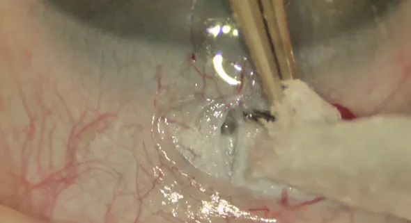
Zeynep Aktas; Gokcen Deniz Gulpinar Ikiz. Current surgical techniques for the management of pediatric glaucoma: A literature review. Frontiers in Ophthalmology. 2023 Mar 22. Figure 2. PMCID: PMC11182127. License: CC BY.
Intraoperative photograph of anterior segment surgery for pediatric glaucoma. It shows manipulation of a tube or instrument in the narrow anterior chamber of a pediatric eye.
Trabeculotomy or goniotomy is often chosen as the initial surgery 3). If these are unsuccessful, trabeculectomy or glaucoma drainage device (GDD) surgery is considered 3).
Many GFCS cases are difficult to treat, and eventually tube shunt surgery with a plate (GDD surgery) may be necessary for long-term intraocular pressure control 1)2)3).
In angle-closure cases, release of pupillary block may be necessary 1). Cyclodestructive procedures may be used as a temporary measure before surgery 3).
In children, attention to side effects is necessary during medical treatment 3).
Alpha-2 agonists: absolute contraindication under 2 years of age, relative contraindication under 7 years of age or body weight under 20 kg
Miotics: risk of pylorospasm and vomiting in infants under 1 month of age
Travoprost: can be used from 2 months of age
QWhy is treatment of GFCS difficult?
A
There are multiple factors. The effect of outflow reconstruction surgery is limited because it often involves developmental abnormalities of the aqueous outflow pathway; the success rate of filtration surgery is low due to the vigorous wound healing response of pediatric ocular tissues; repeated general anesthesia is required; and it must be compatible with amblyopia treatment 3). Collaboration with pediatricians, pediatric ophthalmologists, and orthoptists is essential.
Recent research on GFCS has advanced the elucidation of genetic backgrounds. CYP1B1 gene abnormalities have been identified as a causative gene for PCG, but the full extent of genetic involvement in GFCS has not yet been clarified 4).
Standardization of international childhood glaucoma classification by the Childhood Glaucoma Research Network (CGRN) is progressing, enabling accumulation of epidemiological data and comparison of treatment outcomes for GFCS4).
Risk assessment of glaucoma-related adverse events after cataract surgery associated with persistent fetal vasculature (PFV) is also being conducted 5), and personalized treatment based on risk stratification is expected.
European Glaucoma Society. European Glaucoma Society Terminology and Guidelines for Glaucoma, 5th Edition. Br J Ophthalmol. 2021 Jun;105(Suppl 1):1-169. doi:10.1136/bjophthalmol-2021-egsguidelines. PMID:34675001.
Pazos M, Traverso CE, Viswanathan A; European Glaucoma Society. European Glaucoma Society - Terminology and guidelines for glaucoma, 6th Edition. Br J Ophthalmol. 2025;109(Suppl 1):1-212. doi:10.1136/bjophthalmol-2025-egsguidelines. PMID:41026937.
Freedman SF, Beck AD, Nader DA, et al. International study of childhood glaucoma. Ophthalmol Glaucoma. 2020;3:145-157.
Haider KM, Repka MX, Sutherland DR, Hatt SR, Fallaha N, Kraker RT, Melia BM, Cotter SA, et al. Outcomes and Complications 5 Years After Surgery for Pediatric Cataract Associated With Persistent Fetal Vasculature. American journal of ophthalmology. 2024;260:30-36. doi:10.1016/j.ajo.2023.11.002. PMID:37939986; PMCID:PMC11005992.
Copy the article text and paste it into your preferred AI assistant.
Article copied to clipboard
Open an AI assistant below and paste the copied text into the chat box.