Necrobiotic xanthogranuloma is a type of non-Langerhans cell histiocytosis. It is a chronic granulomatous multisystem disease that mainly affects the skin and tissues around the eye. It was first reported by Kossard and Winkelmann in 1980.1)
It most often begins in the 60s, and there is no difference between men and women.1) Lesions outside the skin can also involve the heart, lungs, bronchi, liver, spleen, oropharynx, and digestive tract.
Its most notable feature is a strong association with blood disorders.
Coexisting paraproteinemia: 95 of 175 cases (55%). IgG-kappa type is the most common subtype1)
Coexisting malignant disease: 19 of 175 cases (11%). Multiple myeloma was the most common, with 12 cases (7%). Hodgkin lymphoma and chronic lymphocytic leukemia have also been reported1)
Overall coexistence of paraproteinemia and/or malignant disease: 114 of 175 cases (65%)1)
Lifetime incidence of hematologic disease: 77–84% of patients with necrobiotic xanthogranuloma develop a hematologic disease at some point in their lives
Progression to multiple myeloma: Cases have been reported up to 6 years after the onset of necrobiotic xanthogranuloma, so long-term follow-up is needed
It is also generally known that multiple myeloma (a neoplastic proliferation of plasma cells) can rarely cause skin xanthomas. A granuloma is a proliferative chronic inflammation characterized by small nodular lesions made up of macrophage-derived epithelioid cells and multinucleated giant cells, and necrobiotic xanthogranuloma is one form of it.
QIf I am diagnosed with necrobiotic xanthogranuloma, will I definitely develop a blood disorder?
A
It is said that 77–84% of patients with necrobiotic xanthogranuloma develop a blood disorder at some point in their lives, so it does not happen in every case, but it is common. Because progression to multiple myeloma has been reported up to 6 years after the onset of necrobiotic xanthogranuloma, regular screening for blood disorders is important.
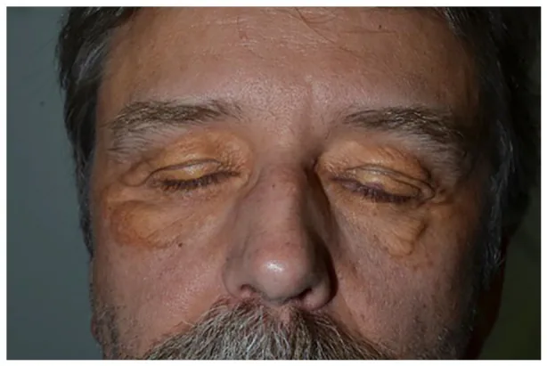
Anne-Sophie Smilga et al. Long-standing necrobiotic xanthogranuloma limited to the skin: A case report. SAGE Open Medical Case Reports. 2021 Nov 9; 9:2050313X211057929. Figure 1. PMCID: PMC8581771. License: CC BY.
Ocular surface complications: subepithelial fibrosis of the cornea and conjunctiva.
QWhat ocular symptoms can occur in necrobiotic xanthogranuloma?
A
About 50% of patients with necrobiotic xanthogranuloma have eye symptoms. In addition to orbital lesions such as orbital masses, proptosis, restrictive strabismus, and ptosis, a variety of findings can occur, including conjunctival lesions, scleritis, keratitis, incomplete eyelid closure (lagophthalmos), and cicatricial ectropion. See the “Clinical findings” section for details.
The direct cause of necrobiotic xanthogranuloma is unknown. A strong association with paraproteinemia is known, but the exact causal relationship has not been clarified.
Age at onset: Average in the 60s, no sex difference1)
Major risk factor: monoclonal gammopathy of undetermined significance (MGUS) and multiple myeloma
Paraprotein hypothesis: monoclonal gammopathy may act as a trigger or cofactor for the giant-cell granulomatous reaction1)
Lipoprotein-binding hypothesis: monoclonal immunoglobulins may bind to lipoproteins and promote uptake by macrophages
Low-HDL hypothesis: patients with necrobiotic xanthogranuloma tend to have low HDL levels (especially HDL-3C), and reduced reverse cholesterol transport from target organs is thought to be involved
Multiple myeloma is a disease caused by the neoplastic proliferation of plasma cells, occurs more often in men over 50, and mainly affects the bones and bone marrow.
The diagnostic criteria proposed by Nelson et al. (2020) are as follows. Apply only when there is no foreign body, infection, or other identifiable cause.
Major criteria (both required)
Skin lesions: presence of papules, patches, or nodules (often yellow to orange).
Pathological findings: palisading granulomas with lymphoplasmacytic infiltration and necrobiotic areas. Cholesterol clefts and giant cells vary by case.
QWhat tests are needed to make a definitive diagnosis of necrobiotic xanthogranuloma?
A
Based on the diagnostic criteria of Nelson et al. (2020), both major criteria (characteristic skin lesions + histopathologic findings) and at least one minor criterion (paraproteinemia or periorbital distribution) must be met. Serum protein electrophoresis is strongly recommended if there is no history of hematologic disease.
There are no consensus guidelines for specific treatment of necrobiotic xanthogranuloma. Randomized controlled trials have not been conducted because it is a rare disease1), and treatment decisions are made by taking into account whether there is an associated malignancy.
The response rates for each drug based on a systematic review (175 cases) are shown below (complete response: CR, partial response: PR).1)
Treatment
Number of cases
Complete response + partial response
Median duration of response
Intravenous immunoglobulin therapy
26 cases
81% (complete 27%, partial 54%)
12 months (range 6-48 months)
Corticosteroids
45 cases
31% (complete 11%, partial 20%)
12 months (range 2-24 months)
Lenalidomide ± steroids
22 cases
50% (complete 18%, partial 32%)
—
Overall, 128 of 175 cases (73%) improved (complete response + partial response), 25 were stable, and 22 cases (13%) progressed. 1)Intravenous immunoglobulin therapy and corticosteroids are recommended as first-line treatment for necrobiotic xanthogranuloma. 1)
For treatment of multiple myeloma, radiation therapy (20–40 Gy) is used for orbital infiltration. Systemic therapy includes melphalan, steroid + melphalan combination therapy, and hematopoietic stem cell transplantation. Molecularly targeted agents used include bortezomib, thalidomide, and the thalidomide derivative lenalidomide (to reduce side effects).
Several drugs have been reported, including cyclosporine, antimetabolites, infliximab, plasmapheresis, alkylating agents (chlorambucil, cyclophosphamide, melphalan), and cladribine.
The recurrence rate after surgical removal is as high as 40%. It is recommended only when severe cicatricial ectropion or lagophthalmos affects vision.
QWhich drug is most effective for treating necrobiotic xanthogranuloma?
A
A systematic review found that intravenous immunoglobulin therapy had the highest response rate (complete response + partial response) at 81%, and it is recommended as a first-line treatment along with corticosteroids.1)Steroid monotherapy has a response rate of only 31%, but combination with intravenous immunoglobulin therapy is standard.
6. Pathophysiology and detailed disease mechanisms
The pathophysiology of necrobiotic xanthogranuloma is not fully understood. Based on its association with paraproteinemia, the following hypotheses have been proposed.
Paraprotein hypothesis: Monoclonal gammopathy acts as a trigger or cofactor for the giant cell granulomatous reaction1)
Lipoprotein-binding hypothesis: Monoclonal immunoglobulin binds to lipoproteins and promotes uptake by macrophages
Low-HDL hypothesis: HDL levels, especially HDL-3C, tend to be low in patients with necrobiotic xanthogranuloma, reducing reverse cholesterol transport from the skin and other target organs
Granulomas are proliferative chronic inflammation characterized by small nodular lesions made up of macrophage-derived epithelioid cells and multinucleated giant cells. The characteristic findings in necrobiotic xanthogranuloma are as follows.
Extent of granuloma: Extends from the dermis to the subcutaneous fat tissue1)
Characteristic giant cells: Touton giant cells and large atypical foreign-body giant cells are frequently seen1)
Cholesterol clefts: A pathological finding characteristic of this disease and important for diagnosis1)
These histological features are thought to reflect a distinctive disease state in which abnormal lipid metabolism and paraproteinemia are combined.
7. Latest research and future prospects (research-stage reports)
Steinhelfer et al. (2022) systematic review (175 cases) is the largest analysis of necrobiotic xanthogranuloma, but it is mostly based on case reports and case series. A fundamental issue is that there is no severity scale for necrobiotic xanthogranuloma, making standardized assessment of treatment response difficult. 1) A prospective randomized controlled trial is needed, but it is considered difficult to carry out because the number of cases is small.
Individual case reports are accumulating for bortezomib, rituximab, adalimumab, mycophenolate mofetil, and clofazimine. 1) Because the number of cases is small, none of these has reached the point of being established as standard treatment.
In addition, a case has been reported in which long-term remission was achieved with combination therapy of monthly intravenous immunoglobulin therapy plus daily thalidomide, cyclophosphamide, and low-dose oral prednisone.