Lacrimal gland tumors are a general term for neoplasms arising from the lacrimal gland within the orbit. The annual incidence is about 1 per million people, accounting for approximately 10% of all orbital space-occupying lesions.
Because the lacrimal gland shares an embryological origin with the salivary glands, the classification system for salivary gland tumors is used for tumor classification.
Lacrimal gland tumors are broadly classified into epithelial tumors and non-epithelial tumors. The table below shows the main classifications and characteristics.
Pleomorphic adenoma: The most common benign tumor, accounting for about 70% of lacrimal gland epithelial tumors. Histologically, it shows a diverse appearance with a mixture of epithelial and stromal components. It is more common in women in their 30s to 40s.
Adenoid cystic carcinoma: The most common epithelial malignant tumor (approximately 60%). More common in men, occurring from adolescence to old age.
Carcinoma ex pleomorphic adenoma: Malignant transformation within a pleomorphic adenoma.
Mucoepidermoid carcinoma: Divided into low-grade and high-grade types.
Primary adenocarcinoma: Very poor prognosis (70% mortality at 3 years).
Lymphomas account for approximately 37% of all malignant lacrimal gland tumors. More common after the age of 60.
Mucosa-associated lymphoid tissue (MALT) lymphoma: Accounts for 70–80% of ocular adnexal lymphomas.
Diffuse large B-cell lymphoma: 10–20%. Rapid progression and poor prognosis.
Follicular lymphoma: Relatively rare.
The annual incidence of ocular adnexal lymphoma is increasing at a rate of 4.5%. 1)
QWhat types of lacrimal gland tumors are there?
A
They are broadly classified into epithelial tumors and non-epithelial tumors. Among epithelial tumors, benign pleomorphic adenoma and malignant adenoid cystic carcinoma are representative, while among non-epithelial tumors, malignant lymphoma (especially MALT lymphoma) accounts for the majority. See the classification table in this section for details.
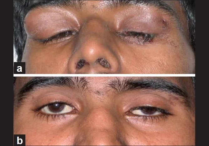
Khanna D, et al. Suppurative dacroadenitis causing ocular sicca syndrome in classic Wegener’s granulomatosis. Indian J Ophthalmol. 2011. Figure 1. PMCID: PMC3116546. License: CC BY.
(a) Bilateral dacryoadenitis with nasal crusting, and (b) disappearance of lacrimal gland tumor after one month of immunosuppressive therapy, although left ptosis is present. Corresponds to lacrimal gland swelling discussed in section “2. Main symptoms and clinical findings.”
Clinical findings vary depending on the type of tumor.
Pleomorphic Adenoma
Ocular deviation: Typical deviation is forward, downward, or toward the nasal side.
Slow growth: Average symptom duration is about 2 years. Characteristically painless.
Palpable subcutaneous mass: A firm, elastic mass is felt on the temporal side of the upper eyelid.
Initial symptoms: About half present with painless unilateral ocular deviation, and the other half with ptosis.
Adenoid Cystic Carcinoma
Rapid growth: Grows faster than pleomorphic adenoma. Symptoms often persist for less than 6 months.
Pain: Pain due to nerve invasion occurs frequently.
Perineural invasion: Occurs in up to 85% of cases. Sensory loss in the first and second divisions of the trigeminal nerve may be observed.
Sudden increase in pain: An important sign suggesting malignant transformation.
Lacrimal gland lymphoma
Age of onset: More common after age 60.
Bilaterality: Approximately 25% of cases are bilateral.
Systemic involvement: Approximately 34% of cases are associated with systemic lymphoma.
Mucosa-associated lymphoid tissue type: Slow, painless course. Large cell type presents with rapid progression and inflammatory findings.
S-shaped deformity (deformity of the upper eyelid) and downward or downward-inward displacement of the eyeball are common findings in lacrimal gland tumors. It may also be accompanied by lower eyelid entropion.
In a case of lacrimal gland mucosa-associated lymphoid tissue lymphoma reported by Zhong et al. in a 60-year-old male, the amount of proptosis was 17 mm on the right and 12 mm on the left, with inferonasal displacement. CT revealed a 3.3 × 2.3 × 2.4 cm mass in the right lacrimal gland. 1)
QWhat are the differences in symptoms between pleomorphic adenoma and adenoid cystic carcinoma?
A
Pleomorphic adenoma is characterized by slow, painless growth, with an average symptom duration of about 2 years. In contrast, adenoid cystic carcinoma grows faster than pleomorphic adenoma and is more likely to be painful. Since perineural invasion occurs in up to 85% of cases, facial sensory abnormalities can also be a clue. However, definitive differentiation between benign and malignant is only possible through histopathological examination.
Adenoid cystic carcinoma: Characterized by MYB-NFIB fusion gene. Involves translocations at 6q22-23 and 9pq23-24.
Malignant transformation of pleomorphic adenoma: Cases of transformation to carcinoma ex pleomorphic adenoma have been reported 5–20 years after excision.
Pathogenesis of mucosa-associated lymphoid tissue lymphoma
Mucosa-associated lymphoid tissue lymphoma develops when chronic antigen stimulation constitutively activates the NF-κB pathway, leading to neoplastic proliferation of B cells. 1)
Helicobacter pylori infection in gastric mucosa-associated lymphoid tissue lymphoma is a classic model of inflammation leading to tumorigenesis. 1)
In lacrimal gland mucosa-associated lymphoid tissue lymphoma, association with Chlamydia psittaci has been reported in over 50% of cases in Italy and South Korea. 1)
Autoimmune diseases (rheumatoid arthritis, Hashimoto thyroiditis, Sjögren syndrome) are also reported to increase the risk of mucosa-associated lymphoid tissue lymphoma. 1)
Interview: Confirm the time of onset, course of worsening, presence of pain, surgical history, and other medical conditions (e.g., autoimmune diseases, malignant tumors).
Palpation: Evaluate the size, hardness, adhesion, and tenderness of the mass. Also check for swelling of regional lymph nodes.
The following table shows the characteristics of major imaging examinations.
Examination
Evaluation Target
Imaging Features of Pleomorphic Adenoma
CT
Bone changes, calcification
Well-defined, spherical to ovoid shape
MRI
Soft tissue
T1 low to iso signal / T2 high signal, moderate enhancement
PET/CT
Evaluation of systemic metastases
—
CT: Useful for evaluating bone changes and calcification. In pleomorphic adenoma, compression and enlargement of the lacrimal fossa, bone sclerosis, and bone thinning are observed. Malignant tumors often have indistinct borders. Tumor size, location, and presence of bone defects are essential for surgical planning.
MRI: Excellent for evaluating soft tissues. Pleomorphic adenoma shows signal intensity equal to or lower than extraocular muscles on T1-weighted images, higher signal intensity than extraocular muscles on T2-weighted images, and moderate contrast enhancement. Large tumors may develop cystic degeneration or calcification internally. MRI is considered the gold standard for diagnosing ocular adnexal lymphoma. 1)
PET/CT and Gallium scintigraphy: Used to evaluate systemic lymphoma or distant metastases.
It is often difficult to differentiate pleomorphic adenoma from malignant epithelial tumors based solely on imaging findings; comprehensive judgment combined with clinical information is necessary.
The biopsy approach varies depending on the suspected disease.
Suspected lymphoma or malignant tumor: Percutaneous incisional biopsy is performed. An incision is made below the lateral eyebrow, and at least approximately 5 mm³ of tumor is resected.
Suspected pleomorphic adenoma: Needle biopsy or incisional biopsy is generally avoided due to the risk of tumor seeding and recurrence from capsular rupture.
Flow cytometry and gene rearrangement tests: Performed to confirm monoclonality in suspected lymphoma cases.
Lacrimal gland cyst: Can be identified by transillumination.
Dacryoadenitis: May be viral or autoimmune. About 30% of lacrimal gland biopsies are diagnosed as idiopathic dacryoadenitis.
IgG4-related ophthalmic disease (Mikulicz disease): Characterized by bilateral gland enlargement. Measurement of IgG4 levels is useful.
Reactive lymphoid hyperplasia: Pathology is essential to differentiate from malignant lymphoma.
Adenoid cystic carcinoma: Presents with pain, rapid growth, and bone destruction.
QIs biopsy necessary even if pleomorphic adenoma is suspected?
A
In principle, biopsy is actively avoided when pleomorphic adenoma is suspected. This is because rupture of the capsule during biopsy can cause tumor cells to seed into surrounding tissues, significantly increasing the recurrence rate. If imaging findings strongly suggest pleomorphic adenoma, direct en bloc excision is performed. On the other hand, if lymphoma or inflammatory disease is suspected, excisional biopsy is indicated.
For pleomorphic adenoma, early surgery is recommended once detected. This is because if the tumor becomes too large, surgery becomes difficult.
En bloc excision is the principle: The tumor must be completely removed as a single piece without rupturing the capsule during the initial surgery. Incomplete resection leads to repeated recurrence.
Needle biopsy and excisional biopsy are contraindicated: They can cause tumor seeding and recurrence.
Pleomorphic adenoma of the eyelid: Can be removed under local anesthesia via an extended blepharoplasty incision.
Orbital pleomorphic adenoma: Requires osteotomy (bone flap osteotomy). Pleomorphic adenomas located in the lacrimal fossa often have the orbital rim bone obstructing removal, making complete excision without osteotomy difficult. The surgical procedure involves osteotomy from the supraorbital notch to the superior border of the zygomatic arch, removal of the tumor, then repositioning the bone and fixing it with periosteal sutures.
Problem of incomplete resection: In cases of incomplete resection, the risk of recurrence increases significantly, and repeated recurrences raise the risk of malignant transformation.
Resectable cases: Aim for complete tumor resection.
Unresectable cases: Perform wide excision (orbital exenteration) plus radiotherapy after incisional biopsy. External beam radiotherapy after tumor debulking is also an option.
Neoadjuvant chemotherapy: Neoadjuvant chemotherapy via the external carotid artery is performed in some cases.
Heavy ion therapy: Currently in clinical trials, being studied as an option with potential for orbital preservation (see “Latest Research” section).
Prognosis varies greatly depending on the histological type.
Pleomorphic adenoma: Prognosis is good as it is a benign tumor, but long-term follow-up is necessary. Malignant transformation to pleomorphic carcinoma may occur 5–20 years after excision. Incomplete resection carries a high risk of recurrence.
Adenoid cystic carcinoma: Mean survival 36 months, 10-year survival 20–30%, poor prognosis. About 50% develop distant metastases (commonly lung and bone). Strong neural and lymphatic invasion, and may invade the brainstem. Follows a high-grade course with bone destruction.
Mucosa-associated lymphoid tissue (MALT) lymphoma: Low metastasis, good response to radiation therapy, relatively good prognosis.
Diffuse large B-cell lymphoma: Prone to metastasis, poor prognosis.
QHow is malignant lymphoma (MALT type) treated?
A
For localized MALT lymphoma, observation or radiation therapy (30 Gy) is first-line. If CD20-positive, rituximab (375 mg/m² weekly for 4 cycles) is effective. For systemic metastasis, chemotherapy such as R-CHOP is indicated. MALT lymphoma responds well to radiation therapy and has a relatively good prognosis.
It is a well-defined tumor surrounded by a pseudocapsule. A biphasic pattern with a mixture of epithelial/myoepithelial cells and mesenchymal components is characteristic, and this histological diversity is the origin of the name “pleomorphic”.
Tumor cells proliferate while forming lumina. The luminal wall has a two-layer structure: the outer layer consists of small cuboidal or spindle-shaped tumor cells with myoepithelial characteristics. These myoepithelial-like cells extend into the stroma and undergo metaplasia to produce mesodermal-derived substances such as mucus (containing glycosaminoglycans) and cartilage.
Outer layer (myoepithelial-like cells): Produce mucus and chondroid matrix.
Inner layer (epithelial cells): Form duct-like structures that secrete glycoproteins.
The pseudocapsule is thin and incomplete, so tumor seeding due to capsular rupture is likely.
Solid and trabecular growth patterns with prominent perineural invasion are characteristic. Tumor cells are small with chromatin-rich nuclei. Perineural invasion can be confirmed by neurofilament staining.
Histological subtypes are classified into five types.
Cribriform type: The most common subtype. Characterized by sieve-like cavity formation.
Solid type
Sclerosing type
Comedocarcinomatous type
Tubular type
Polymorphous adenocarcinoma and mucoepidermoid carcinoma
It shows diffuse proliferation of lymphoid cells, and monoclonality (monoclonal B-cell proliferation) can be confirmed by immunostaining and Southern blotting.
In the case reported by Zhong et al., immunohistochemical staining showed positivity for CD20, CD79a, PAX5, and CD10, and negativity for BCL2, with positivity for BCL6. The Ki67 proliferation index was approximately 60% in germinal centers and about 15% in plasmacytoid areas. 1)
MYB-NFIB fusion gene: Fusion of transcription factors MYB and NFIB leads to overexpression of MYB target genes, causing abnormal cell proliferation, survival, and differentiation.
Chromosomal translocation: Involves translocations at 6q22-23 and 9pq23-24.
Molecular mechanisms of mucosa-associated lymphoid tissue lymphoma
Chronic antigenic stimulation (infection with Chlamydia psittaci, autoimmune reactions) persistently activates B-cell receptor signaling. 1)
This constitutively activates the NF-κB pathway, promoting proliferation and survival of neoplastic B cells. 1)Helicobacter pylori-associated gastric MALT lymphoma is a classic model where tumor regression has been confirmed after eradication of the infection. 1)
7. Latest research and future perspectives (reports at research stage)
Heavy particle (carbon ion) therapy is undergoing clinical trials primarily for adenoid cystic carcinoma. Compared to conventional external beam radiation, it offers higher dose concentration due to the Bragg peak, allowing high-dose irradiation while preserving the orbit. Currently, it is at the clinical trial stage and has not been established as standard treatment.
Fluorescence in situ hybridization (FISH) is positioned as an important tool for molecular genetic confirmation in the diagnosis of mucosa-associated lymphoid tissue lymphoma. 1)
Monitoring of immunoglobulin heavy chain (IGH) and immunoglobulin kappa chain (IGK) gene rearrangements is expected to enable assessment of treatment response and detection of minimal residual disease. 1)
The ratio of regulatory T cells (Treg) to helper T17 cells has been suggested to be associated with tumor outcomes, and elucidation of the immune microenvironment is expected to lead to the discovery of new therapeutic targets. 1)
Zhong Q, Yan Y, Li S.. Mucosa-associated lymphoid tissue lymphoma of the lacrimal gland: A case report and literature review. Medicine (Baltimore). 2024;103(21):e38303. doi:10.1097/md.0000000000038303. PMID:38787969; PMCID:PMC11124633.