Migraine is a type of vascular headache, a paroxysmal headache disorder caused by dilation of branches of the external carotid artery. It is characterized by recurrent throbbing headaches, broadly divided into those with and without aura. Scintillating scotoma is a typical visual aura preceding migraine, classically described as zigzag patterns of light caused by vasospasm leading to transient ischemia of the visual cortex in the occipital lobe. Currently, cortical spreading depression (CSD) is understood as the main mechanism (see §6).
It lasts 4 to 72 hours, accompanied by nausea, photophobia, phonophobia, and visual aura. It is classified as episodic (less than 15 days per month) and chronic (15 or more days per month for more than 3 months).
Hemiplegic migraine: Aura with reversible motor weakness. Prevalence 0.01%. Classified into familial (CACNA1A, ATP1A2, SCN1A gene mutations) and sporadic forms2).
Recurrent Painful Ophthalmoplegic Neuropathy (RPON), formerly known as “ophthalmoplegic migraine,” was renamed in ICHD-3. The reason for the renaming is that it is more characteristic of a demyelinating inflammatory neuropathy than migraine.
It is a rare disorder characterized by recurrent attacks of headache and ipsilateral ophthalmoplegia, with an incidence of 0.7 per million people. It primarily occurs in children under 10 years of age, with a median age of onset reported as 8 years. Adult onset is also recognized; in a pooled analysis of 165 cases, the mean age of onset was 22.1 years, and 34.2% of cases occurred at age 18 or older 6). The oculomotor nerve (III) is most commonly affected, followed by the abducens nerve (VI) and trochlear nerve (IV).
QWhy do eye symptoms occur with migraine?
A
Cortical spreading depression (CSD) and involvement of the trigeminovascular system are the main causes. CSD is a wave of neuronal depolarization that begins in the visual cortex of the occipital lobe, causing scintillating scotoma. Additionally, activation of the trigeminovascular system releases inflammatory substances such as CGRP and substance P, leading to vasodilation and neurogenic inflammation, which cause photophobia and headache.
Photophobia (light sensitivity): The most common ocular symptom in migraine patients. Light worsens the headache. Almost always bilateral.
Visual aura (scintillating scotoma): Zigzag, sawtooth, or crescent-shaped flickering lights that expand and move from the center to the periphery of the visual field. They disappear within 20–30 minutes and are often followed by a throbbing headache. Characteristically binocular and homonymous. Visual aura is present in over 90% of cases with aura.
Attack frequency: Varies from once a month to twice a week among patients. The pain peaks within 1–2 hours and is often accompanied by nausea and vomiting.
Visual persistence (palinopsia): Afterimages of objects that have disappeared from the visual field remain. Commonly seen in migraine with aura.
Visual snow: Small particles like TV static spread across the entire visual field. It can persist for years. 60% of patients with visual snow syndrome (VSS) have comorbid migraine.
Alice in Wonderland Syndrome (AIWS): Micropsia, macropsia, and distortion of one’s own body parts. Common in males aged 5–14 and females aged 16–18.
QWhat is the difference between scintillating scotoma and transient monocular blindness?
A
The main differences are in the nature of the visual symptoms, laterality, and duration. Scintillating scotoma is a positive symptom (zigzag lights) that is binocular and lasts 20–30 minutes (up to 60 minutes). Transient monocular blindness is a negative symptom (darkening or graying) that is monocular and resolves quickly within 1–5 minutes (up to 10 minutes). Transient monocular blindness may be caused by embolism of the carotid or ophthalmic artery and should not be overlooked.
Clinical findings by subtype presenting with ocular signs are shown below.
Retinal Migraine
Monocular vision loss: Reversible monocular visual impairment or loss.
Scotoma: C-shaped, colored, scintillating, expanding scotoma. Diagnosis requires at least 2 episodes.
Exclude TMVL: Always differentiate from serious causes (arteritis, embolism).
Hemiplegic Migraine
Motor weakness: Unilateral reversible motor paralysis occurs as an aura.
Various auras: Accompanied by visual, sensory, and language symptoms. Each symptom lasts 5 to 60 minutes.
ICHD-3 criteria: Develops over ≥5 minutes, with headache occurring with the aura or within 60 minutes2).
RPON (Ophthalmoplegic Neuropathy)
Third cranial nerve palsy: Oculomotor nerve most commonly affected (in children). Ptosis, eye movement disorder, mydriasis.
MRI findings: Localized contrast enhancement and thickening of the oculomotor nerve in the acute phase. A literature review of 52 cases reported MRI third nerve enhancement in 75% and nerve swelling in 76%6).
Recurrence: Two or more attacks are required for a definitive diagnosis. In some cases, recovery becomes incomplete with repeated attacks9).
Basilar-type migraine
Brainstem symptoms: accompanied by dizziness, dysarthria, ataxia, tinnitus, and hearing loss.
Diplopia: may be accompanied by bilateral sensory abnormalities or altered consciousness as a typical aura.
Syncope: transient loss of consciousness may occur in severe cases.
Headache: Unilateral, most intense around the orbit or behind the eye. Often persists for several days to about a week, tending to be longer than typical migraine (less than 72 hours). It does not necessarily have to be migraine-like (ICHD-III criteria). Up to one-third of patients do not have classic migraine features.
Associated symptoms: Photophobia 65%, nausea 66%, vomiting 69% (systematic review by Gelfand et al.).
Diplopia and ptosis: Appear with the onset of ophthalmoplegia.
Absence of aura: Visual, sensory, or speech auras have not been reported, an important distinction from typical migraine.
There is a time lag from headache onset to the appearance of ophthalmoplegia, ranging from immediately to up to 14 days. Ophthalmoplegia usually resolves spontaneously within 2 weeks to 3 months, but incomplete recovery may occur after recurrent attacks. In cases with long-term recurrence, 54% develop persistent ocular motor palsy 9).
Oculomotor Nerve (III)
Ptosis: Drooping due to paralysis of the levator palpebrae superioris muscle.
Restricted eye movement: Limitation of adduction, elevation, and depression.
Mydriasis and decreased light reflex: Pupillary fibers are often involved. However, in a review of 39 cases by McMillan et al., the pupil was spared in 23%.
Abducens Nerve (VI)
Abducens palsy: Esotropia due to limited abduction.
In adults, this is the cranial nerve most commonly affected in RPON.
Trochlear Nerve (IV)
Limitation of depression and intorsion: Vertical diplopia is the main complaint.
Compensatory head tilt to the opposite side is observed. Relatively rare involvement.
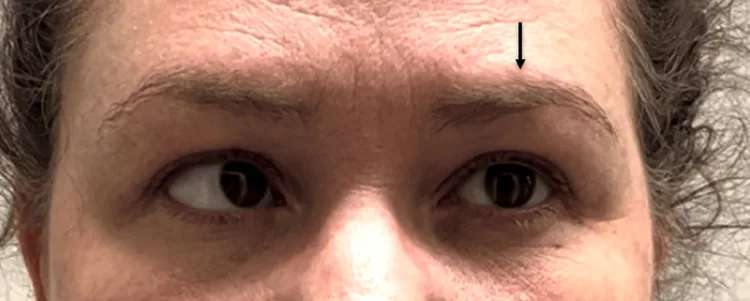
Attempting leftward gaze. The left eye remains fixed in neutral due to left cranial nerve VI (abducens) palsy, while the right eye fully adducts. Image obtained and shown with patient consent. Arrow added for emphasis
In typical migraine, ophthalmic examinations (including visual field tests) are usually normal. During a migraine attack, OCTA (optical coherence tomography angiography) may show a marked decrease in choroidal vascular density and enlargement of the foveal avascular zone (FAZ).
QHow long does it take for ophthalmoplegia to resolve?
A
It usually resolves spontaneously within 2 weeks to 3 months, but in some cases recovery becomes incomplete after repeated attacks. In long-standing recurrent cases, persistent ocular motor palsy has been reported in 54% of patients 9). Although it improves spontaneously after onset, the duration may lengthen with each attack and become permanent in some cases.
Genetic factors: In familial hemiplegic migraine, mutations in CACNA1A, ATP1A2, and SCN1A genes (autosomal dominant inheritance) 2).
Stroke risk: Migraine with aura is a risk factor for stroke. The risk is particularly high in women under 45 years old who use oral contraceptives and smoke.
History of migraine: A personal or family history of migraine is frequently observed.
Preceding infection: Cases following viral gastroenteritis 10) and after tympanitis 8) have been reported. Infection may trigger immune-mediated neuropathy.
Pregnancy: Onset at 19 weeks of pregnancy and spontaneous remission on the fifth day postpartum have been reported, suggesting an association with hormonal and physiological changes 7).
Regional differences and genetic diversity: An association with abnormal hemoglobin has also been reported in Nigerian populations.
QWhat foods or medications can worsen migraines?
A
Food triggers include red wine, beer, chocolate, aged cheese, MSG (monosodium glutamate), and aspartame. Medications that may trigger or worsen migraines include oral contraceptives, estrogen therapy, nasal decongestants, opioids, and SSRIs. Since triggers vary greatly among individuals, self-monitoring with a headache diary is important.
Migraine diagnosis is based on medical history, physical examination, and ICHD-3 criteria. Imaging is not necessary for typical symptoms. Diagnostic tools include ID-Migraine, VARS, MIDAS questionnaire, and MSQ 2.11).
If migraine is suspected, first exclude organic diseases such as cerebrovascular disorders or neoplastic conditions using CT or MRI. Next, prescribe a drug that suppresses vasospasm and confirm the diagnosis by checking whether the attack can be aborted with this medication.
Indications for imaging (brain MRI/CT) are limited to the following cases:
Unexplained abnormal neurological findings
New onset or progressive worsening after age 40
Suspicion of migrainous cerebral infarction
Sudden severe headache (requires exclusion of subarachnoid hemorrhage)
MRI is the most important diagnostic test. Characteristic findings include local contrast enhancement and thickening of the cisternal segment of the oculomotor nerve. In a literature review of 52 cases, MRI showed third nerve enhancement in 75% and nerve swelling in 76% 10). However, MRI may be normal in 25–81% of acute cases. Contrast enhancement usually resolves within 7–9 weeks but may persist for 2–4 years in some cases.
A complete MRI evaluation including TOF-MRA is recommended from the first attack 5). In oculomotor palsy involving pupillary function, MRA is particularly important to exclude posterior cerebral artery (PCA) aneurysm.
Headache secondary to trauma, infection, or congenital disease: Exclusion of secondary headache is necessary.
Clinical tests include cerebrospinal fluid analysis (usually normal; to differentiate from malignancy and MS) 5), and blood tests to rule out diabetes, infection, inflammation, and autoimmune diseases.
QIs it possible to have RPON even if MRI is normal?
A
Yes. In 25–81% of acute cases, MRI findings may be normal 5). In RPON with normal MRI, a strategy of observation with NSAIDs and assessment of response has been reported. However, in recurrent or persistent cases, repeat MRI is essential to exclude other organic lesions.
Treatment for migraine includes preventive medications, drugs used during the aura phase, and medications for acute attacks, which are used appropriately.
For mild attacks, NSAIDs, dihydroergotamine mesylate, and oral triptans are effective. For severe attacks, oral triptans are used.
NSAIDs (ibuprofen, aspirin): Side effects are milder than triptans.
Triptans (sumatriptan 25–100 mg): More effective than NSAIDs. Combination of triptan + NSAIDs has a higher 2-hour pain-free rate than either alone 1). Sumatriptan is available as oral, subcutaneous injection, and nasal spray.
Dihydroergotamine mesylate (Dihydergot): Effective for mild to moderate attacks.
Lasmiditan: 5-HT1F receptor agonist. A new drug for acute treatment 1).
When aura occurs, dihydroergotamine mesylate should be used early. Since administration in the early stage of a migraine attack enhances efficacy, prompt administration upon sensing aura is important.
No standardized treatment guidelines exist. For acute attacks, NSAIDs and non-narcotic analgesics are used. For prevention, beta-blockers, calcium channel blockers, tricyclic antidepressants, and antiepileptic drugs are used. For frequent attacks, long-acting verapamil or lamotrigine may be used.
Although symptoms improve spontaneously after onset, some attacks become prolonged and permanent, so aggressive anti-inflammatory therapy is recommended.
Corticosteroids are the most common acute treatment. Pooled analysis showed rapid improvement in 96.2% of 76 patients treated with steroids 6). For inflammatory lesions of oculomotor nerve palsy, prednisolone 50–60 mg/day is given for the first 3 days, then tapered carefully to avoid relapse.
The main steroid regimens are shown below.
Regimen
Dosage and administration
Report
Methylprednisolone (pediatric)
25–30 mg/kg/day (max 1 g/day) × 5 days
Frattini 20235), Nandana 20219)
Methylprednisolone (adults)
250 mg 4 times/day for 3 days → prednisone 60 mg tapering
For RPON with normal MRI, one option is to observe with NSAIDs (ibuprofen 10 mg/kg/dose, 3 times/day). There are reports of complete recovery within 48 hours with ibuprofen 5).
QDoes the preventive drug topiramate have ocular side effects?
A
Topiramate is widely used as a migraine preventive, but it can cause acute angle closure (TiAAC) around 2 weeks after starting treatment. Ciliochoroidal effusion causes forward movement of the lens-iris diaphragm, leading to a rapid increase in intraocular pressure. If eye pain, vision loss, or blurred vision occur, promptly consult an ophthalmologist and contact the prescribing physician4).
QAre there preventive medications for RPON?
A
Calcium channel blockers such as flunarizine and verapamil, beta-blockers such as propranolol, and antiepileptic drugs such as valproic acid, gabapentin, and topiramate are used as preventive medications. Since there is a 30% risk of permanent sequelae, it is important to consider active preventive therapy in cases of frequent recurrence 6).
The basic mechanism of migraine is stimulation of the meninges, blood vessels, and trigeminal nerve territory, triggered by dilation of branches of the external carotid artery 1).
Triggers such as stress, food, and hormones → dysregulation of brainstem vascular control → peripheral vasodilation → stretch signals to trigeminal neurons → production of inflammatory and vasoactive substances such as CGRP and interleukins → further dilation and increased vascular permeability → tissue edema forms a cascade 1).
The neurotransmitters involved are substance P, nitric oxide, and CGRP. The periaqueductal gray (PAG), locus coeruleus (LC), and raphe nuclei (DRN) are brain regions implicated in the pathophysiology of migraine.
Cortical Spreading Depression (CSD) and Visual Aura
Classically, it was explained that cerebral vasospasm leads to transient ischemia in the occipital visual cortex, causing scintillating scotoma. Currently, cortical spreading depression (CSD) is widely supported as the main mechanism of visual aura 2).
A wave of neuronal depolarization originating in the occipital region propagates forward. Depolarization increases potassium concentration, and the release of excitatory amino acids further enhances spreading. CSD causing temporary dysfunction of the occipital visual cortex corresponds to the occurrence of scintillating scotoma.
Basilar-type migraine: CSD in the brainstem is involved.
Retinal migraine: CSD in the retina (although there is also much evidence suggesting a cortical mechanism, and some point out that “retinal migraine” is a misnomer).
Hemiplegic migraine: Vasogenic leakage from pial vessels stimulates the trigeminovascular system, causing hemiplegia as an aura 2).
AIWS and visual snow: AIWS results from transient ischemia of the visual pathway. Visual snow involves hypermetabolism of the secondary visual cortex (lingual gyrus, Brodmann area 19).
Several hypotheses have been proposed regarding the pathogenesis of RPON, and debate continues.
Demyelination/Inflammation Theory (Mainstream)
Lance & Zagami: Recurrent demyelinating neuropathy/neuritis theory. MRI oculomotor nerve enhancement is cited as evidence.
Carlow hypothesis: A neuropeptide cascade from the ophthalmic branch of the trigeminal nerve induces aseptic inflammatory vascular reaction in the circle of Willis. Repeated demyelination and remyelination lead to onion bulb proliferation of Schwann cells8).
Ischemia Theory
Ambrosetto et al.: Vasospasm of vasa nervorum during migraine → reversible ischemic disruption of the blood-nerve barrier → vasogenic edema (explains MRI enhancement and thickening).
Vijayan et al.: ICA wall edema → occlusion of small vessel openings → watershed infarction-type injury. Shin et al.’s SPECT study confirmed reversible ipsilateral ischemia in the PCA perforator territory.
Neurovascular compression theory
Bui et al. reported a case of a 13-year-old boy in whom MRA confirmed compression of the oculomotor nerve exit zone by a sharp loop of the left posterior cerebral artery (PCA)10).
Arguments against the compression theory: (1) pupillary sparing in 23% of cases, (2) no vascular stenosis demonstrated on carotid angiography during attacks, and (3) recovery from RPON is slower than after decompression.
Among these hypotheses, the demyelination/inflammation theory is currently mainstream. Similarities to the relapsing-remitting patterns of CIDP, MS, and MOGAD have been noted, but CSF studies are usually normal, and direct evidence of viral infection or immune-mediated neuropathy has not been obtained.
During spontaneous migraine attacks, OCTA shows a marked decrease in choroidal vascular density and enlargement of the FAZ. These findings suggest that choroidal circulation is more vulnerable than retinal circulation during attacks.
Three genes are known to be associated with familial hemiplegic migraine: CACNA1A (calcium channel), ATP1A2 (Na/K pump), and SCN1A (sodium channel)2). Mutations in the TREK gene (two-pore potassium channel) are involved in impaired regulation of resting membrane potential and neuronal hyperexcitability1).
7. Latest Research and Future Perspectives (Research-stage Reports)
Fremanezumab, erenumab, and galcanezumab are FDA-approved preventive medications for chronic migraine1).
A meta-analysis of anti-CGRP mAbs showed a significant improvement in the 50% responder rate, with reductions in monthly migraine days and acute medication use. There is broad agreement that their benefit-risk profile is superior to propranolol and topiramate 1).
Galcanezumab, with a regimen of a 240 mg subcutaneous loading dose followed by 120 mg monthly for 5 months, improved headache severity, frequency, and duration. Injection site itching and rash have been reported as main side effects 1).
Ubrogepant was FDA-approved in 2019 as an oral acute treatment. It can be used with or without aura 1). Atogepant is being developed as an oral preventive medication.
Tonabersat and Treatments Targeting Potassium Channels
Tonabersat is a novel benzopyran molecule that inhibits glial-neuronal gap junction communication and suppresses CSD. A 39-patient RCT (Goadsby et al. 2009) showed efficacy in preventing migraine with aura, but it is currently not FDA-approved 1).
Mutations in two-pore potassium channels (TREK) cause impaired regulation of resting membrane potential, leading to neuronal hyperexcitability. Therapeutic research targeting TREK activation/inhibition is ongoing 1).
Takemoto et al. (2021) reported a 14-year-old girl with steroid-induced intraocular pressure elevation whose diplopia markedly improved within days and ptosis and ocular movement disorder completely resolved after administration of 0.5% timolol maleate eye drops 8). It is speculated that topical timolol may have exerted an effect on RPON through neuropeptide-mediated inhibition of the trigeminal vascular system. Beta-blocker eye drops may be effective even without elevated intraocular pressure, and further exploratory studies are expected.
Koo et al. (2024) proposed revised diagnostic criteria based on Liu et al.’s pooled analysis: “at least two unilateral headache attacks (almost simultaneous with or up to 15 days before ophthalmoplegia)” plus “clinical findings of cranial nerve palsy or MRI enhancement” 6). Discussions continue to include adult-onset and atypical cases.
Castillo-Guerrero et al. (2024) reported a case of internal ophthalmoplegic migraine during pregnancy (bilateral mydriasis without external ophthalmoplegia) 7). Spontaneous remission on the fifth postpartum day was confirmed, suggesting that hormonal and physiological changes may be involved in the onset.
An association with anti-GQ1b antibody syndrome has been reported, and in recurrent cases that respond rapidly to steroids, testing for atypical anti-GQ1b antibody syndrome is recommended 5). Additionally, evaluation of neurovascular compression using high-resolution MRA is becoming increasingly important in the diagnosis of pediatric RPON, and compression of the oculomotor nerve exit by the PCA has been confirmed in some cases 10).
Okobi OE, Boms MG, Ijeh JC, Eboigbe SE, Alex KB, Adejola AA, et al. Migraine and Current Pharmacologic Management. Cureus. 2022;14(10):e29833. doi:10.7759/cureus.29833. PMID:36337819; PMCID:PMC9626373.
Kana T, Mehjabeen S, Patel N, et al. Sporadic Hemiplegic Migraine. Cureus. 2023;15(5):e38930.
Falsaperla R, Presti S, Lo Bianco M, Catanzaro S, Marino S, Ruggieri M. Diagnostic controversies in recurrent painful ophthalmoplegic neuropathy: single case report with a systematic review. Italian journal of pediatrics. 2022;48(1):82. doi:10.1186/s13052-022-01274-x. PMID:35659705; PMCID:PMC9164546.
Al Owaifeer AM, AlSultan ZM, Badawi AH. Topiramate-induced acute angle closure: A systematic review of case reports and case series. Indian journal of ophthalmology. 2022;70(5):1491-1501. doi:10.4103/ijo.IJO_2134_21. PMID:35502014; PMCID:PMC9333044.
Koo H, Tsai K, Lee C, Mustafa I. A Case of Adult-Onset Recurrent Painful Ophthalmoplegic Neuropathy With Bilateral Ophthalmoplegia. Cureus. 2024;16(2):e54683. doi:10.7759/cureus.54683. PMID:38523969; PMCID:PMC10960561.
Castillo-Guerrero B, Londoño-Juliao G, Pianetta Y, Gutiérrez-Rey M, Zuñiga BJ, Pestana G, et al. Internal Ophthalmoplegic Migraine During Pregnancy: A Clinical Case. Neurology international. 2024;16(6):1779-1787. doi:10.3390/neurolint16060128. PMID:39728754; PMCID:PMC11677585.
Takemoto D, Ohkubo S, Udagawa S, Kuroda M, Sugiyama K. A Case of Recurrent Painful Ophthalmoplegic Neuropathy Successfully Treated with Beta-blocker Eye Drop Instillation. Neuro-ophthalmology (Aeolus Press). 2021;45(5):320-323. doi:10.1080/01658107.2020.1791190. PMID:34483410; PMCID:PMC8409776.
Nandana J, Nair SS, Girdhar S, Sundaram S. Recurrent painful ophthalmoplegic neuropathy: a cause for recurrent third nerve palsy in a child. BMJ case reports. 2021;14(11). doi:10.1136/bcr-2021-246179. PMID:34764123; PMCID:PMC8587473.
Bui LT, Mainali G, Naik S, Cockroft K, Thamburaj K. Role of Neurovascular Compression of Oculomotor Nerve in Ophthalmoplegic Migraine. Cureus. 2022;14(3):e22919. doi:10.7759/cureus.22919. PMID:35399433; PMCID:PMC8985736.
Mrabet S, Nasri A, Kessentini N, Ben Djebara M, Gargouri-Berrechid A, Kacem I, et al. Recurrent painful ophthalmoplegic neuropathy revealing oculomotor nerve schwannoma. La Tunisie medicale. 2021;99(8):919-923. PMID:35261021; PMCID:PMC9003581.
Copy the article text and paste it into your preferred AI assistant.
Article copied to clipboard
Open an AI assistant below and paste the copied text into the chat box.