Recurrent Painful Ophthalmoplegic Neuropathy (RPON) is a rare disorder characterized by recurrent episodes of unilateral headache and ipsilateral oculomotor nerve palsy. Previously called “ophthalmoplegic migraine” (OM), it was renamed in the International Classification of Headache Disorders, 3rd edition (ICHD-3) because its features are more consistent with a demyelinating inflammatory neuropathy than with migraine.
The incidence is 0.7 per million people. It mainly occurs in children under 10 years of age, with a median age of onset reported as 8 years. Unlike common migraines, it tends to be slightly more common in boys during childhood. Adult onset is also observed; in a pooled analysis by Liu et al. (165 cases), the mean age of onset was 22.1 years, with 34.2% of cases occurring at age 18 or older2).
The most commonly affected cranial nerve is the oculomotor nerve (III), followed by the abducens nerve (VI) and the trochlear nerve (IV). In children, oculomotor nerve palsy is most common, but in adults, abducens nerve palsy tends to be the most frequent 5).
Historically, Notta first reported it as “periodic ophthalmoplegia” in 1854, and Charcot used the term “ophthalmoplegic migraine.” Harold Wolff formally described it in 1939 3).
QCan adults get this disease?
A
Adult onset has also been reported; in Liu et al.’s analysis, 34.2% of cases occurred at age 18 or older 2). However, it is mainly seen in children under 10 years old and is relatively rare in adults.
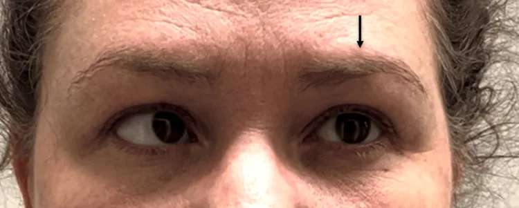
Nonpharmacologic Management of Recurrent Painful Ophthalmoplegic Neuropathy: A Case Report. Cureus. 2025 May 28; 17(5):e84387. Figure 2. PMCID: PMC12178448. License: CC BY.
Attempting leftward gaze. The left eye remains fixed in neutral due to left cranial nerve VI (abducens) palsy, while the right eye fully adducts. Image obtained and shown with patient consent. Arrow added for emphasis
Headache: Unilateral, most intense around the orbit or behind the eye. It often persists for several days to about a week, tending to be longer than typical migraine (less than 72 hours).
Nature of headache: Not necessarily migraine-like (ICHD-III criteria). Up to one-third of patients do not have classic migraine features.
Associated symptoms: Photophobia 65%, nausea 66%, vomiting 69% (systematic review by Gelfand et al.).
Diplopia: Appears with the onset of ophthalmoplegia.
Ptosis: Appears when the oculomotor nerve is affected.
Absence of aura: No visual, sensory, or speech aura is reported, which is an important distinguishing feature from typical migraine.
There is a time lag from headache onset to the appearance of ophthalmoplegia, ranging from immediately to up to 14 days. Ophthalmoplegia usually resolves spontaneously within 2 weeks to 3 months, but some cases show incomplete recovery after recurrent attacks. In long-term recurrent cases, 54% develop persistent ocular motor palsy 5).
Findings by affected cranial nerve are shown below.
Oculomotor Nerve (III)
Ptosis: Drooping due to paralysis of the levator palpebrae superioris muscle.
Restricted eye movement: Limitations in adduction, supraduction, and infraduction.
Mydriasis and decreased light reflex: Pupillary fibers are often involved. However, in a review of 39 cases by McMillian et al., 23% had pupil sparing.
Abducens nerve (CN VI)
Abducens palsy: Esotropia due to limited abduction.
In adults, it is the most commonly affected cranial nerve in RPON.
Trochlear nerve (CN IV)
Limited downward and inward rotation: Vertical diplopia is the main complaint.
Compensatory head tilt to the opposite side is observed. Relatively rare involvement.
QHow long does it take for ocular muscle palsy to resolve?
A
It usually resolves spontaneously within 2 weeks to 3 months, but in some cases recovery becomes incomplete after repeated attacks. In long-term recurrent cases, persistent ocular motor palsy has been reported in 54%5). Although it improves spontaneously after onset, the duration may lengthen with each attack and become permanent in some cases.
The exact cause of RPON is unknown, but the following associations have been noted.
History of migraine: A high frequency of migraine history in the patient or family members.
Genetic predisposition: Familial clustering suggests a possible genetic involvement.
Prior infection: Cases have been reported following viral gastroenteritis6) and tympanitis4). Infection may trigger immune-mediated neuropathy.
Pregnancy: Onset at 19 weeks of pregnancy and spontaneous remission on the 5th postpartum day have been reported, suggesting an association with hormonal and physiological changes3).
Regional differences and genetic diversity: An association with abnormal hemoglobin has also been reported in Nigerian populations.
MRI is the most important diagnostic test. Characteristic findings are focal contrast enhancement and thickening of the oculomotor nerve in the cisternal segment. A literature review of 52 cases reported MRI enhancement of the third nerve in 75% and nerve swelling in 76% 6). However, MRI may be normal in 25–81% of acute cases. Contrast enhancement usually resolves within 7–9 weeks but may persist for 2–4 years in some cases.
Complete MRI evaluation including TOF-MRA is recommended from the first attack 1). In oculomotor palsy involving pupillary function, MRA is especially important to exclude posterior cerebral artery (PCA) aneurysm.
QIs RPON possible even if MRI is normal?
A
In 25–81% of acute cases, MRI findings may be normal 1). For RPON with normal MRI, a method of first observing the response to NSAIDs has also been reported. However, in recurrent or persistent cases, it is important to repeat MRI to rule out other organic lesions.
Ophthalmoplegia resolves within 72 hours (RPON lasts ≥2 weeks)
Clinical tests include cerebrospinal fluid analysis (usually normal; to differentiate from malignancy and MS) 1), and blood tests to rule out diabetes, infection, inflammation, and autoimmune disease. For differential diagnosis of oculomotor nerve palsy, imaging (MRI/MRA), Tensilon test, ice pack test, and anti-AChR antibody measurement are also important.
Although it improves spontaneously after onset, active anti-inflammatory therapy is recommended because the duration of each attack may become longer and some cases become persistent.
Corticosteroids are the most common acute phase treatment. In a pooled analysis by Liu et al., 96.2% of 76 patients treated with steroids showed rapid improvement 2). Active anti-inflammatory therapy is recommended. For inflammatory lesions of oculomotor nerve palsy, prednisolone 50–60 mg/day is first administered for 3 days, then tapered with caution for relapse during early dose reduction.
For RPON with normal MRI, observation with NSAIDs (ibuprofen 10 mg/kg/dose three times daily) is an option. Complete recovery within 48 hours with ibuprofen has been reported1). Also, more than 50% of patients recover completely within 72 hours without treatment1).
The indication for preventive therapy is typical migraine with frequent attacks; otherwise, evidence is insufficient. Since 30% of patients have a risk of permanent neurological sequelae (residual cranial nerve vulnerability, pupillary dysfunction), the importance of early treatment and prevention has been emphasized2).
Calcium channel blockers such as flunarizine and verapamil, beta-blockers such as propranolol, and antiepileptic drugs such as valproic acid, gabapentin, and topiramate are used as preventive medications. Since there is a 30% risk of permanent sequelae, it is important to consider aggressive preventive therapy in cases of frequent recurrence 2).
Several hypotheses have been proposed regarding the pathogenesis of RPON, and debate continues.
Demyelination/Inflammation Theory (Mainstream)
Lance & Zagami: Recurrent demyelinating neuropathy/neuritis theory. MRI enhancement of the oculomotor nerve is considered evidence.
Carlow hypothesis: A neuropeptide cascade from the ophthalmic branch of the trigeminal nerve induces an aseptic inflammatory vascular reaction in the circle of Willis. Repeated demyelination and remyelination lead to onion bulb proliferation of Schwann cells4).
Ischemic theory
Ambrosetto et al.: Vasospasm of the vasa nervorum during migraine → reversible ischemic disruption of the blood-nerve barrier → vasogenic edema (explaining MRI contrast enhancement and thickening).
Vijayan et al.: ICA wall edema → occlusion of small vessel openings → border zone infarct-like injury. Shin et al.’s SPECT study confirmed reversible ipsilateral ischemia in the territory of the posterior communicating artery perforators.
Neurovascular compression theory
Bui et al.: Reported a 13-year-old boy with compression of the oculomotor nerve exit zone by a sharp loop of the left posterior cerebral artery (PCA) on MRA6).
Objections to the compression theory: (1) Pupil sparing in 23% of cases, (2) Carotid angiography during attacks does not demonstrate vascular stenosis, (3) Recovery from RPON is slower than after decompression.
Among these hypotheses, the demyelination/inflammation theory is currently mainstream. Similarities to relapsing-remitting patterns of CIDP, MS, and MOGAD have been pointed out, but CSF tests are usually normal, and direct evidence of viral infection or immune-mediated neuropathy has not been obtained. While THS presents similar symptoms, RPON differs in that there is no mass-like contrast enhancement in the cavernous sinus.
7. Latest Research and Future Perspectives (Research-stage Reports)
Takemoto et al. (2021) reported a 14-year-old girl with steroid-induced intraocular pressure elevation whose diplopia markedly improved within days and ptosis and ocular movement disorder completely resolved after administration of 0.5% timolol maleate eye drops 4). It is hypothesized that topical timolol may have exerted effects on RPON through neuropeptide-mediated inhibition of the trigeminovascular system (Carlow hypothesis). Beta-blocker eye drops may be effective even without elevated intraocular pressure, and further exploratory studies are expected.
Based on a pooled analysis by Liu et al., Koo et al. (2024) proposed revised diagnostic criteria for ICHD-III: “at least two unilateral headache attacks (occurring almost simultaneously with or up to 15 days before ophthalmoplegia)” plus “clinical signs of cranial nerve palsy or MRI enhancement” 2). Discussions continue toward including adult-onset and atypical cases.
Castillo-Guerrero et al. (2024) reported a case of internal ophthalmoplegic migraine during pregnancy (bilateral mydriasis without external ophthalmoplegia) 3). Spontaneous remission on postpartum day 5 was confirmed, suggesting that hormonal and physiological changes may contribute to onset. Memantine (NMDA receptor antagonist) 10 mg/day was used experimentally for migraine prophylaxis, but evidence for use during pregnancy is limited.
An association with anti-GQ1b antibody syndrome has been reported, and in recurrent cases that respond rapidly to steroids, screening for atypical anti-GQ1b antibody syndrome is recommended 1). Additionally, high-resolution MRA evaluation of neurovascular compression is gaining importance in pediatric OM diagnosis, and compression of the oculomotor nerve exit by the PCA has been confirmed in some cases 6).
Frattini D, Iodice A, Spagnoli C, et al. Tolosa-Hunt syndrome and recurrent painful ophthalmoplegic neuropathy, case reports: what to do and when? Ital J Pediatr. 2023;49:157.
Koo H, Tsai K, Lee C, Mustafa I. A Case of Adult-Onset Recurrent Painful Ophthalmoplegic Neuropathy With Bilateral Ophthalmoplegia. Cureus. 2024;16(2):e54683. doi:10.7759/cureus.54683. PMID:38523969; PMCID:PMC10960561.
Castillo-Guerrero B, Londoño-Juliao G, Pianetta Y, Gutiérrez-Rey M, Zuñiga BJ, Pestana G, et al. Internal Ophthalmoplegic Migraine During Pregnancy: A Clinical Case. Neurology international. 2024;16(6):1779-1787. doi:10.3390/neurolint16060128. PMID:39728754; PMCID:PMC11677585.
Takemoto D, Ohkubo S, Udagawa S, Kuroda M, Sugiyama K. A Case of Recurrent Painful Ophthalmoplegic Neuropathy Successfully Treated with Beta-blocker Eye Drop Instillation. Neuro-ophthalmology (Aeolus Press). 2021;45(5):320-323. doi:10.1080/01658107.2020.1791190. PMID:34483410; PMCID:PMC8409776.
Nandana J, Nair SS, Girdhar S, Sundaram S. Recurrent painful ophthalmoplegic neuropathy: a cause for recurrent third nerve palsy in a child. BMJ case reports. 2021;14(11). doi:10.1136/bcr-2021-246179. PMID:34764123; PMCID:PMC8587473.
Bui LT, Mainali G, Naik S, Cockroft K, Thamburaj K. Role of Neurovascular Compression of Oculomotor Nerve in Ophthalmoplegic Migraine. Cureus. 2022;14(3):e22919. doi:10.7759/cureus.22919. PMID:35399433; PMCID:PMC8985736.
Mrabet S, Nasri A, Kessentini N, Ben Djebara M, Gargouri-Berrechid A, Kacem I, et al. Recurrent painful ophthalmoplegic neuropathy revealing oculomotor nerve schwannoma. La Tunisie medicale. 2021;99(8):919-923. PMID:35261021; PMCID:PMC9003581.
Copy the article text and paste it into your preferred AI assistant.
Article copied to clipboard
Open an AI assistant below and paste the copied text into the chat box.