Rhino-orbital-cerebral mucormycosis (ROCM) is an invasive fungal infection caused by fungi of the order Mucorales, particularly Rhizopus oryzae. It is a fatal disease that rapidly progresses from the paranasal sinuses to the orbit and brain, and was previously called orbital zygomycosis. It was first described by J.E. Gregory in 19431).
Rhizopus species account for approximately 85–90% of rhinocerebral cases. The estimated annual incidence is 1.7 per million people8), and in India, the incidence is reported to be 80 times higher than in developed countries9). In Europe, the incidence is reported as 0.2–3 per million4). The overall mortality rate exceeds 50%, reaching 79% without treatment and approximately 40.5% with treatment. Central nervous system involvement occurs in 33–49% of diabetic patients with poor glycemic control8).
Although rare, the mortality rate is high, with a reported mortality rate of 94% for the invasive form of fungal sinusitis. Since the COVID-19 pandemic, cases have increased rapidly, especially in India6).
QHow often does rhino-orbital-cerebral mucormycosis occur?
A
The estimated annual incidence is 1.7 per million people8). In India, the incidence is reported to be 80 times higher than in developed countries, showing significant regional variation. The risk of developing the disease is markedly increased in patient populations with diabetes, hematologic malignancies, or immunodeficiency.
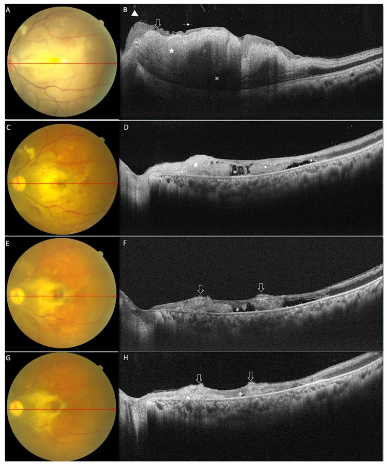
Spectral Domain Optical Coherence Tomography Findings in Vision-Threatening Rhino-Orbital Cerebral Mucor Mycosis—A Prospective Analysis. Diagnostics (Basel). 2022 Dec 8; 12(12):3098. Figure 1. PMCID: PMC9777225. License: CC BY.
Serial fundus photographs and SD-OCT images of a representative case (case 5), at baseline (A) Fundus photographs showing diffuse retinal whiting with papilla-macular fold, boxcar segmentation of the vessel, absence of cherry red spot (B) The corresponding SD-OCT line scan shows diffuse retinal thickening, increased inner retinal hyperreflectivity (white star) with shadowing effect on the outer retina (white asterisk). Along with these retinal folds, inner limiting membrane (ILM) detachment (empty arrow), vitreous haze and vitreous cells (arrowhead) are also seen. At three weeks (C) fundus photographs show a reduction in the area of diffuse retinal whiting, with few cotton wool spots and few hemorrhages. (D) On SD-OCT disruption of all retinal layers (white star) along with optically empty
In all cases of COVID-19-associated ROCM, CRAO, proptosis, and complete ophthalmoplegia were observed, and the prognosis was extremely poor (all cases died) 3).
Fungi of the order Mucorales, especially Rhizopus oryzae, are the main causative organisms. They are fast-growing, aseptate filamentous fungi, widely distributed in tropical and subtropical regions. They enter the body through inhalation of spores.
QDoes having COVID-19 make you more likely to develop mucormycosis?
A
Cases of ROCM after COVID-19 infection have increased rapidly worldwide, with 2,826 cases reported in India in 2021 alone 6). The cytokine storm, immune abnormalities, steroid use, and hyperglycemia caused by COVID-19 are thought to synergistically promote fungal growth.
CT: Useful for evaluating bone erosion of the sinus walls and opacification of the sinus cavities. The most commonly involved sites are the maxillary and ethmoid sinuses. Contrast-enhanced CT is preferred. Many cases show no abnormalities in the early stages, so repeated imaging is important.
Biopsy and culture are the gold standard for definitive diagnosis. Histopathology uses H&E staining, PAS staining, and GMS staining, and characteristic findings include broad, ribbon-like, nonseptate hyphae branching at right angles (90 degrees), vascular invasion, thrombosis, and necrosis 2). Species identification by culture is also important for selecting treatment, but the culture positivity rate is low. Definitive diagnosis is made by demonstrating the organism through histopathological examination or culture.
DNA sequences of Rhizopus species can be detected in cerebrospinal fluid. Positivity was reported in all 7 cases (all cultures were negative), and it is particularly useful when conventional methods are negative.
The characteristics of the diagnostic methods are shown below.
Test method
Features
Cautions
Biopsy + culture
Gold standard for definitive diagnosis
Low culture positivity rate
mNGS (cerebrospinal fluid)
Detectable even in culture-negative cases
Requires specialized facility
β-D-glucan and GM
Routine fungal markers
Low sensitivity to Mucorales, unsuitable for diagnosis
The basic approach is a combination of antifungal drugs and surgical debridement, and treatment in an internal medicine department capable of systemic management is desirable. Since antifungal drug penetration into necrotic tissue is poor, combination with surgical treatment is essential.
Duration: 3–36 months (based on clinical and imaging improvement)
Amphotericin B deoxycholate has significantly higher nephrotoxicity; liposomal formulations are preferred.
Posaconazole: Step-down or salvage therapy. Response rate 60–70%2).
Isavuconazole: Effective as alternative therapy with good tolerability7). 200 mg loading dose, then 200 mg/day (oral available).
Combination therapy: Amphotericin plus caspofungin has been reported to have better outcomes than monotherapy, but echinocandins have low in vitro activity against Mucorales, and strong evidence is lacking2).
Surgical debridement of necrotic tissue is essential. Resection should be performed until normal bleeding is obtained, and pathological confirmation of the resection margin is strongly recommended. Functional endoscopic sinus surgery (FESS) is the standard procedure, and multiple surgeries may be required 4). In cases with extensive orbital involvement, orbital exenteration with bone removal may be necessary.
The mortality rate for antifungal therapy combined with surgery is 18.5%, whereas it is significantly higher at 60% for antifungal therapy alone 2).
Blood glucose control in diabetes and correction of diabetic ketoacidosis7)
Correction of immunosuppressive state (reduction of immunosuppressive drugs as much as possible)
QHow long does treatment take?
A
The duration of liposomal amphotericin B administration ranges widely from 3 to 36 months, and is individually adjusted based on clinical and imaging improvement 2). Severe cases or those with brain involvement often require longer administration. Surgical debridement may also need to be performed multiple times.
Inhalation of spores leads to proliferation within the paranasal sinuses, reaching the orbit via direct invasion or through the nasolacrimal duct. From the orbit, invasion into the brain occurs through the orbital apex, cavernous sinus, cribriform plate, and blood vessels.
The main mechanism of spread is angioinvasion, penetrating the endothelial cells of blood vessel walls and extracellular matrix proteins. GRP78 (glucose-regulated protein) is involved in this penetration process. It progresses through the pathway of angioinvasion → thrombosis → ischemia → avascular necrosis, resulting in necrosis without congestion.
Under acidic conditions, iron is released from transferrin, and Mucorales fungi utilize the free iron to proliferate rapidly. The same mechanism occurs in iron overload states (hemochromatosis, frequent blood transfusions, deferoxamine therapy).
Cytokine storm (elevation of IL-1, IL-2, IL-6, TNF-α, etc.), decreased IFN-γ expression in CD4+ T cells, use of steroids and immunomodulators, and the overlap of hypoxic environment + hyperglycemia + acidic environment + high iron levels provide an ideal environment for fungal spore germination7).
Yang et al. (2026) reported 7 cases of ROCM presenting with cerebral infarction as the initial symptom5). In all cases, Rhizopus species were detected by mNGS of cerebrospinal fluid, but cultures were negative in all cases. The median time to diagnosis was 5 days, and the only survivor was diagnosed within 2 days (mortality rate 85.7%). mNGS is a promising early diagnostic tool for ROCM, especially valuable when conventional cultures and serum markers are negative.
QIn what situations is mNGS testing useful?
A
mNGS (metagenomic next-generation sequencing) is useful when β-D-glucan and galactomannan have poor sensitivity and cultures fail to identify the causative organism. In atypical ROCM presenting with cerebral infarction as the initial symptom, cerebrospinal fluid mNGS may be the only diagnostic method5).
Rapid Increase in COVID-19-Associated ROCM and Countermeasures
In response to the global surge in ROCM associated with COVID-19, Ostovan et al. (2021) reported that all patients with a history of mechanical ventilation died 6). In India, Sen et al. (2021) reported 2,826 cases of COVID-19-associated ROCM, highlighting it as a significant new complication of the pandemic.
Al Reesi et al. (2023) reported a case of a child with acute chronic kidney disease and malnutrition who achieved cure with aggressive treatment including liposomal amphotericin B (5→9 mg/kg/day) plus posaconazole plus multiple surgeries 2). Early diagnosis within 24 hours and aggressive treatment are considered key to favorable outcomes.
Benlamkaddem S, Zdaik G, Doughmi D, Bennis A, Chraibi F, Berdai MA, et al. Rhino-Orbital Cerebral Mucormycosis: A Fatal Evolution. Cureus. 2023;15(4):e37837. doi:10.7759/cureus.37837. PMID:37214071; PMCID:PMC10198304.
Al Reesi M, Al Muqbali T, Al Ajmi A, et al. Successful Management of Rhino-Orbital-Cerebral Mucormycosis in a Child with Acute-on-Chronic Kidney Disease and Malnutrition. Sultan Qaboos Univ Med J. 2023.
Kamath GM, Jeganathan S, Salim S, Antony RM, Kamath AR, Hiran H. Case series of central retinal artery occlusion in COVID-19-associated rhino-orbital-cerebral mucormycosis. Indian journal of ophthalmology. 2023;71(7):2904-2906. doi:10.4103/IJO.IJO_3123_22. PMID:37417144; PMCID:PMC10491029.
Siriwardena P, Wariyapperuma U, Nanayakkara P, Jayawardena N, Mendis D, Bahar M, et al. Rhino-orbital-cerebral mucormycosis in acute myeloid leukemia patients: a case series from Sri Lanka. BMC infectious diseases. 2024;24(1):1465. doi:10.1186/s12879-024-10334-y. PMID:39725915; PMCID:PMC11670406.
Yang F, Yang C, Li H, Zhang X, Ding X, Zhang S. Metagenomic next-generation sequencing in diagnosing rhino-orbital-cerebral mucormycosis presenting as cerebral Infarction: a case series and diagnostic analysis of seven patients. Frontiers in fungal biology. 2026;7:1751546. doi:10.3389/ffunb.2026.1751546. PMID:41659795; PMCID:PMC12873710.
Ostovan VR, Rezapanah S, Behzadi Z, Hosseini L, Jahangiri R, Anbardar MH, et al. Coronavirus disease (COVID-19) complicated by rhino-orbital-cerebral mucormycosis presenting with neurovascular thrombosis: a case report and review of literature. Journal of neurovirology. 2021;27(4):644-649. doi:10.1007/s13365-021-00996-8. PMID:34342852; PMCID:PMC8330178.
Ponce-Rosas L, Gonzales-Zamora J, Diaz-Reyes N, et al. Rhino-Orbital-Cerebral Mucormycosis in a Post-COVID-19 Patient from Peru. Case Rep Infect Dis. 2022.
Alanazi RF, Almalki A, Alkhaibary A, AlSufiani F, Aloraidi A. Rhino-Orbital-Cerebral Mucormycosis: A Rare Complication of Uncontrolled Diabetes. Case reports in surgery. 2022;2022:6535588. doi:10.1155/2022/6535588. PMID:36245688; PMCID:PMC9556216.
Mokhtar EA, Fatima Q, Akbar S, Equbal S, Salahudeen A. Rhino-Orbital Cerebral Mucormycosis Causing Temporomandibular Joint Ankylosis: A Case Series of Two Patients. Cureus. 2023;15(2):e35194. doi:10.7759/cureus.35194. PMID:36960262; PMCID:PMC10030649.
Copy the article text and paste it into your preferred AI assistant.
Article copied to clipboard
Open an AI assistant below and paste the copied text into the chat box.