Cherry-red spot (CRS) is one of the fundus findings at the posterior pole. It is named because the foveola appears red against the surrounding whitened retina, resembling a cherry. In Japanese, it is also called “sakura-jitsu kouhan” (cherry fruit red spot).
This finding was first described in 1887 by Bernard Sachs as a fundus finding in “familial amaurotic idiocy” (now Tay-Sachs disease). It is now widely recognized as a clinically important sign appearing in various diseases such as CRAO and various lysosomal storage diseases.
The incidence of CRAO is about 1 per 100,000 people 1). 1-2% of CRAO cases are bilateral, suggesting vasculitis such as giant cell arteritis (GCA) 10). Also, about 60% of ophthalmic artery occlusion (OAO) cases show CRS9).
Appearance varies by race: bright red in Caucasians, but brown or blackish in non-Caucasians. This is due to differences in choroidal pigmentation. The alternative term “perifoveal white patch” has also been proposed.
2. Diseases and causes presenting with cherry-red spot
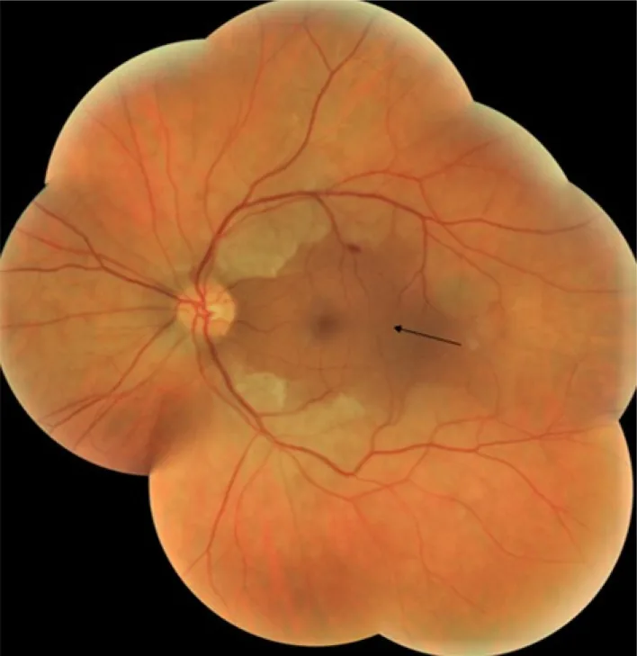
Anis Mahmoud; Fatma Abid; Molka Khairallah; Fatma Sakji; Hassen Ibn Hadj Amor; Hala Attia. Case Report: Central retinal artery occlusion following sildenafil intake. F1000Res. 2022 Jun 1; 11:600 Figure 1. PMCID: PMC9490277. License: CC BY.
Baseline composite fundus photograph of the left eye showing retinal whitening except for the cilioretinal artery distribution area (black arrow).
CRS is not a single disease but a clinical sign resulting from multiple different conditions. The causes are broadly classified into four groups: “vascular,” “metabolic storage diseases,” “drugs/toxins,” and “trauma/other.”
Vascular (CRAO)
Non-arteritic CRAO: Accounts for approximately 95% of all CRAO cases. Types of emboli: cholesterol emboli 74%, platelet-fibrin emboli 15.5%, calcium emboli 10.5% 2). Carotid artery disease is present in over 70% of cases (EAGLE study) 1).
Arteritic CRAO: Giant cell arteritis (GCA) is the main cause. Bilateral CRAO must always rule out GCA 10).
CRAO due to infective endocarditis: Caused by septic emboli. Embolic complications occur in 22–50% of IE patients 9).
Metabolic Storage Diseases
Tay–Sachs disease: Hexosaminidase A deficiency. CRS frequency 75–90%.
GM1 gangliosidosis type 1: β-galactosidase deficiency. CRS frequency approximately 50%.
Sialidosis type 1: Neuraminidase (NEU1) deficiency. CRS appears in almost all cases 3)5). However, absence of CRS has been reported in biallelic mild NEU1 mutations 5).
Drugs/Toxins
PDE5 inhibitors (sildenafil, etc.): There are case reports of CRAO after overdose of sildenafil 100 mg × 2 tablets 6). The FDA has accumulated many reports of ocular vascular occlusion related to PDE5 inhibitors.
Others: Quinine poisoning, carbon monoxide (CO) poisoning, etc.
Trauma/Other
Commotio retinae: Retinal whitening at the posterior pole after ocular contusion, presenting a pseudo-CRS.
CRAO after COVID-19 vaccine: The first worldwide report of CRAO after Covaxin (inactivated vaccine from India) was documented 8).
Sickle cell trait + diabetes: Sickling due to oxidative stress can cause CRAO1).
The frequency of CRS appearance by metabolic storage disease is summarized.
In children, lysosomal storage diseases are the main cause. In Tay-Sachs disease, CRS appears in 75-90% of cases 5). When CRS is seen in children along with neurological symptoms such as myoclonus, ataxia, and seizures, evaluation for metabolic storage disease is necessary.
Cherry-red spot (CRS): Contrast between retinal whitening (opacity) at the posterior pole and the red color of the fovea. In CRAO, it is seen in about 90% of cases at initial examination.
Retinal arteriolar narrowing: Narrowing of retinal arteries 1)4)8).
Boxcarring: Interruption of blood flow in retinal arteries, appearing as a beaded pattern 4)10).
Visible emboli (Hollenhorst plaque): Cholesterol emboli may be visible (20-40%).
Roth spots: White-centered hemorrhages that appear in cases of infective endocarditis2).
OCT: In the acute phase, hyperreflectivity and thickening of the inner retinal layers1)2)6)8). In the chronic phase, thinning and atrophy of the inner retinal layers1)7).
Fundus examination: Confirmation of cherry-red spot under dilated pupil and evaluation of retinal arteries. Check for the presence of emboli.
RAPD assessment: Confirmation of afferent pupillary defect using the swinging flashlight test.
Visual acuity and visual field testing: Assessment of the extent and severity of occlusion.
Imaging
OCT: Evaluation of acute hyperreflectivity and chronic thinning of the inner retinal layers. In lysosomal storage diseases, early changes may be detected earlier than with standard fundus examination5).
OCTA: Non-invasive assessment of areas of blood flow loss6).
FA: Confirmation of delayed arterial filling pattern1). Fundus autofluorescence (FAF) is useful for evaluating lysosomal storage diseases5).
Systemic Examination
Cardiac Ultrasound (TEE): Transesophageal echocardiography has a sensitivity of 91–100%. It is superior to transthoracic echocardiography (TTE), which has a sensitivity of 44–63%2).
Carotid Ultrasound: Evaluation of carotid artery stenosis and plaque4).
CTA/MRI (Brain): Confirmation of concurrent cerebral infarction4).
Coagulation Tests and Hb Electrophoresis: Evaluation of thrombophilia and sickle cell trait1).
If metabolic storage disease is suspected:
NEU1 gene testing: Confirms the genotype of sialidosis5).
Enzyme activity measurement: Measures the activity of each lysosomal enzyme to determine the type of storage disease3).
QIs a cherry-red spot an emergency?
A
In CRAO, it is an emergency equivalent to stroke. Irreversible retinal damage occurs approximately 4 hours after onset7), and 12.9% of inpatients have concurrent stroke and 3.7% have myocardial infarction4). Immediate emergency ophthalmology and neurology consultation is necessary upon noticing symptoms.
Currently, there is no established standard treatment with proven efficacy for CRAO9). The following treatments may be attempted, but evidence for each is limited.
Spontaneous improvement after CRAO onset occurs in only about 22% of untreated cases 7). The time until irreversible retinal damage occurs is estimated to be about 4 hours 7).
Treatments attempted in the acute phase are summarized below.
Regarding vitrectomy, a surgical technique has been reported that combines lowering intraocular pressure to less than 3 mmHg with an increase in arterial pressure via intravenous injection of 0.1 mg adrenaline. Okonkwo et al. performed this procedure in two cases of non-arteritic CRAO and reported improvement in visual acuity to 6/60 and 6/36+1, respectively 7).
Ischemia-reperfusion injury (IRI) is thought to occur approximately 7 days after CRAO7).
Secondary prevention:
Antiplatelet agents and statins: Aspirin 150 mg/day plus atorvastatin 20 mg at night is used for secondary prevention 4).
CRAO due to infective endocarditis: Base treatment on 6 weeks of antibiotics; consider valve replacement if vegetations exceed 15 mm 2)9).
Metabolic storage diseases: Clonazepam, sodium valproate, and levetiracetam are used to manage myoclonus 5).
QDoes treatment for CRAO improve vision?
A
There is no established treatment, and about 22% of untreated cases improve spontaneously 7)9). Case reports of vitrectomy have recorded recovery to visual acuity of 6/60 or 6/36+1 7), but no large-scale comparative trials exist. Early response after onset and management of systemic complications are important.
6. Pathophysiology and detailed mechanism of onset
In the macula, the ganglion cell layer forms a thick structure with multiple layers. When blood flow to the inner retina is interrupted due to occlusion of the central retinal artery (CRA), ganglion cells rapidly become ischemic and swell, causing the posterior pole retina to appear milky white.
In the foveola, there is no ganglion cell layer. Therefore, the foveola receives direct nutrition from the choroidal circulation and remains transparent. As a result, the red color of the choroid becomes visible in the foveola against the surrounding opaque retina—this is the cause of the cherry-red spot.
In 15–25% of eyes, a cilioretinal artery is present. Since this artery branches from the choroidal circulation independently of the central retinal artery, central vision may be preserved in cases of CRAO10).
The retina can tolerate ischemia for about 97 minutes, but irreversible damage occurs after 4 hours 7). Ischemia-reperfusion injury (IRI) is thought to occur approximately 7 days after CRAO7).
Due to lysosomal enzyme deficiencies, undegraded lipids (such as gangliosides and sialylated glycoproteins) accumulate within ganglion cells. This accumulation causes swelling of the cell bodies, leading to opacification of the ganglion cell layer in the macula. Since the foveola lacks ganglion cells, a cherry-red spot appears through the same mechanism as in CRAO.
In sialidosis, mutations in the NEU1 gene cause neuraminidase deficiency, leading to accumulation of sialylated glycoproteins 3)5). The cherry-red spot may appear late as the accumulation increases, and conversely, it may become less distinct as ganglion cell death progresses 5). If mild NEU1 mutations are present on both alleles, the cherry-red spot may be absent 5).
PDE5 inhibitors increase cGMP, which may lead to decreased cerebral blood flow (CBF) due to systemic hypotension 6). Furthermore, long-term elevation of cGMP is thought to alter endothelial permeability and promote platelet adhesion and thrombus formation 6).
QCan cherry-red spots be absent in sialidosis?
A
It has been reported that when mild NEU1 gene mutations are present on both alleles, the cherry-red spot may be absent 5). Even in cases where the cherry-red spot is indistinct, OCT or fundus autofluorescence (FAF) may detect abnormalities more sensitively 5). The absence of a cherry-red spot should not rule out a storage disease.
QDoes the appearance of the cherry-red spot differ by race?
A
In Caucasians, it appears as a bright red color, but in non-Caucasians (e.g., Asians, Africans), it may appear brown to black. This is due to differences in choroidal pigmentation, and caution is needed when interpreting the finding.
7. Latest Research and Future Perspectives (Research-Stage Reports)
Surgical treatment of CRAO combining vitrectomy with intraocular pressure and arterial pressure manipulation has been reported.
Okonkwo et al. (2021) performed vitrectomy (lowering intraocular pressure to <3 mmHg plus intravenous adrenaline 0.1 mg to increase arterial pressure) in two cases of non-arteritic CRAO and reported improvement in visual acuity to 6/60 and 6/36+1, respectively 7). It is thought that the shorter the time after onset, the higher the possibility of visual function recovery.
The AHA/ASA 2021 guidelines recommend treating CRAO as equivalent to stroke and performing neurological evaluation within 72 hours.
Zhong Yang et al. (2024) reported a case of CRAO complicated by concurrent cerebral infarction, showing a post-admission stroke risk of 12.9%, myocardial infarction risk of 3.7%, and total cardiovascular event risk of 19% in CRAO patients 4). Collaboration between stroke teams and ophthalmology is required.
Thakar et al. (2022) reported the world’s first case of CRAO after Covaxin (inactivated COVID-19 vaccine) vaccination 8). It is attracting attention as a vaccine-related vascular complication, and elucidating the causal relationship is a future challenge.
Sahoo et al. (2023) identified a novel NEU1 mutation (c.544T>A, p.Y182N) in sialidosis and reported a clinical picture characterized by cherry-red spot and myoclonus 3). Neeraja et al. (2021) reported a case of type 1 sialidosis with a mild NEU1 mutation lacking a cherry-red spot, showing that OCT and autofluorescence are useful for diagnosis 5). Genotype-phenotype correlation analysis is progressing.
Semidey VA, Magliyah MS, Alali N, Hashem F, ALBalawi HB. Central Retinal Artery Occlusion in a Young Patient With a Hidden Unusual Sickle Cell Trait. Cureus. 2023;15(2):e34865. doi:10.7759/cureus.34865. PMID:36923174; PMCID:PMC10010448.
Harshvardhan Chawla, Jonah S. Goldblatt, John E. Morgan, Bruce A. Barron, Aravinda K. Rao, Maria A. Reinoso. Central Retinal Artery Occlusion with Concomitant Intracranial Hemorrhage Secondary to Streptococcus Gordonii Endocarditis. Case Reports in Ophthalmological Medicine. 2023;2023:1-5. doi:10.1155/2023/9268480.
Sahoo LK, Mohapatra S, Jena RK, et al. Novel NEU1 variant in sialidosis presenting with cherry-red spot. Neurology. 2023;101(19):861-862.
Zhong Yang L, Ngoo QZ, Nilamani V, Sudarno R. A Triggering Event of Central Retinal Artery Occlusion With Concurrent Ischemic Stroke. Cureus. 2024;16(2):e53577. doi:10.7759/cureus.53577. PMID:38445140; PMCID:PMC10914528.
Neeraja K, Rukmini AV, Pal PK, et al. Sialidosis type I without cherry red spot: a diagnostic challenge. J Mov Disord. 2021;14(1):65-69.
Abrishami M, Hosseini SM, Mohseni H, Razavi M, Ghaffarian Mashhadi Nejad A, Baghi Yazdi M, et al. Central Retinal Artery Occlusion Associated with Sildenafil Overdose. Case reports in ophthalmological medicine. 2021;2021:2006271. doi:10.1155/2021/2006271. PMID:34527380; PMCID:PMC8437635.
Ogugua Ndubuisi Okonkwo, Adekunle Olubola Hassan, Toyin Akanbi, Victor Chukwuebuka Umeh, Oladapo Oluwadamilola Ogunbekun. Vitrectomy and manipulation of intraocular and arterial pressures for the treatment of nonarteritic central retinal artery occlusion. Taiwan Journal of Ophthalmology. 2021;11(3):305-311. doi:10.4103/tjo.tjo_51_20.
Thakar M, Bhattacharya S. Central retinal artery occlusion after vaccination with whole virion inactivated SARSCoV- 2 vaccine Covaxin. Indian journal of ophthalmology. 2022;70(10):3716-3718. doi:10.4103/ijo.IJO_1148_22. PMID:36190081; PMCID:PMC9789834.
Mohamed M, Alsawidi K, Elhousseini Z, et al. Bilateral central retinal artery occlusion in infective endocarditis. J VitreoRetinal Dis. 2021;5(3):261-265.
American Academy of Ophthalmology. Retinal and ophthalmic artery occlusions preferred practice pattern. AAO; 2024.
Copy the article text and paste it into your preferred AI assistant.
Article copied to clipboard
Open an AI assistant below and paste the copied text into the chat box.