Traumatic optic neuropathy (TON) is a condition in which a strong blow to the forehead or frontal region, especially the eyebrow area, causes transmitted force (force that propagates rather than direct impact) to act on the optic canal, resulting in contusion of the optic nerve. Importantly, it is not necessarily associated with optic canal fracture; severe optic nerve damage can occur even in cases without fracture.
Typically, it develops after blunt trauma to the superolateral eyebrow area, so most cases show subcutaneous hemorrhage or contusion lacerations on the lateral side of the eyebrow.
TON is reported to occur in 0.5–5% of all head injuries. Common mechanisms include traffic accidents, sports injuries, and falls, with a typical pattern of blunt trauma to the superolateral brow. Only a minority of cases show optic canal deformity on plain X-ray or CT.
In the 2021 Beirut port explosion, 39 patients (48 eyes) were evaluated ophthalmologically. Orbital fractures were found in 14 eyes (29.2%), open globe injuries in 10 eyes (20.8%), and 53.8% required surgical intervention 1). Explosive trauma can also cause TON (see “Causes and Risk Factors” section for blast-induced traumatic optic neuropathy).
QHow is traumatic optic neuropathy different from optic nerve head avulsion?
A
Traumatic optic neuropathy is a condition in which the optic nerve is damaged at the optic canal due to indirect force from blunt trauma to the brow area. In contrast, optic nerve head avulsion is a severe injury where the optic nerve is physically torn at the level of the lamina cribrosa, and the avulsion site can be seen on fundus examination immediately after injury. A key distinguishing feature is that the fundus is usually normal immediately after injury in traumatic optic neuropathy.
GCC thickness becomes thinner and falls below normal range
6–8 weeks after injury and later
Progressive optic atrophy and optic disc pallor
GCC thickness stabilizes around 30–50 days
Relative afferent pupillary defect (RAPD) is the most important objective finding in unilateral or asymmetric bilateral cases, and is confirmed as Marcus-Gunn pupil (positive swinging flashlight test in the affected eye).
QCan traumatic optic neuropathy be ruled out even if the fundus is normal immediately after injury?
A
It cannot be ruled out. Immediately after injury, the fundus is often normal. Optic atrophy and optic disc pallor appear 6–8 weeks after injury, and thinning of GCC thickness on OCT is observed from about 2 weeks after injury. A normal fundus immediately after injury is not grounds for excluding the diagnosis; functional assessments such as RAPD (swinging flashlight test) are important.
Blunt trauma to the superolateral brow is the most common mechanism of injury. The impact propagates through the optic canal, causing vasogenic edema within the optic nerve parenchyma (see Pathophysiology section for details on pathophysiology).
Main causes:
Traffic accidents: Most common. Contact with windshield, airbag, or steering wheel.
Sports injuries: Collision with racket, ball, or ground.
Falls: Impact of face or forehead with the ground.
Assault: Direct blunt force to the face from fists or blunt objects.
Shock waves from blast overpressure propagate through ocular structures to the optic nerve, causing shear forces and stress that damage optic nerve fibers. It is characterized by the absence of penetrating injury or significant blunt trauma, and optic neuropathy can occur even without visible external signs of injury.
Military personnel, emergency responders, and civilians exposed to explosives are at high risk.
65–68% of blast-injured soldiers with traumatic brain injury (TBI) report visual problems.
Animal models have confirmed a dose-response relationship between total blast exposure and the degree of optic nerve degeneration.
Elevation of IL-1α and IL-1β in the optic nerve and retina has been demonstrated in animal models.
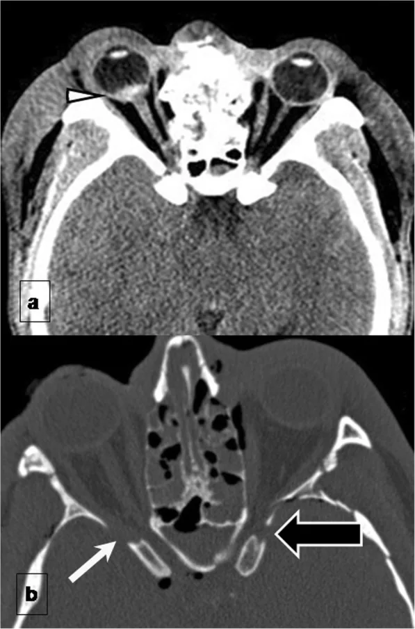
Gómez Roselló E, Quiles Granado AM, Artajona Garcia M, et al. Facial fractures: classification and highlights for a useful report. Insights Imaging. 2020;11(1):49. Figure 2. PMID: 32193796; PMCID: PMC7082488; DOI: 10.1186/s13244-020-00847-w. License: CC BY.
Axial non-contrast CT of the orbit shows (a) hematoma at the posterior pole of the globe extending to the optic disc (arrowhead) and (b) bone fragment within the muscle cone causing optic nerve injury (black arrow: bone fragment, white arrow: optic nerve transection). This corresponds to the diagnostic findings of orbital fracture and optic nerve injury on CT discussed in the section “4. Diagnosis and Examination Methods.”
The most important test for diagnosing optic neuropathy is the swinging flashlight test. In the affected eye, the pupil dilates when light is shone, confirming a positive relative afferent pupillary defect (RAPD) (Marcus Gunn pupil). Even if visual acuity and fundus findings are good, this finding indicates the presence of optic nerve injury.
The following tests are used in combination for evaluation.
QIs it possible to have traumatic optic neuropathy even with good visual acuity?
A
Yes. In traumatic optic neuropathy, even if visual acuity is relatively preserved, visual field abnormalities, decreased contrast sensitivity, color vision abnormalities, and positive RAPD may be present. Evaluation using only high-contrast visual acuity risks missing the impairment. Assessment with RAPD (swinging flashlight test) is essential.
Early diagnosis (within 24–48 hours after injury) and prompt and appropriate reduction of optic nerve parenchymal edema significantly affect prognosis.
Pharmacotherapy
Steroid pulse therapy: Intravenous administration of prednisone equivalent 1,000 mg/day for 2–3 days is a standard option.
High-dose steroid therapy: Systemic administration of prednisolone equivalent 80–100 mg/day. Used as an alternative similar to pulse therapy.
Hyperosmotic agents: Intravenous infusion of Glycerol® or D-mannitol 300–500 mL for 3–7 days to reduce optic nerve parenchymal edema.
Tapering: Gradually reduce steroids while monitoring visual progress.
Surgical Treatment
Optic canal decompression: There is much controversy regarding the indications for open surgery. Many believe that reducing edema within the optic nerve parenchyma is difficult to achieve surgically, except in cases where the optic nerve is clearly compromised due to marked deformation of the optic canal or significant displacement of bone fragments.
Transnasal endoscopic approach: In recent years, a minimally invasive transnasal endoscopic approach has become feasible.
Limited indications: Cases with marked deformation of the optic canal or significant displacement of bone fragments are considered appropriate for surgery.
Severe cases where loss of light perception does not recover quickly: These cases tend to respond poorly to treatment.
Stabilization of visual function: Even in cases that have progressed several weeks after injury, aggressive pharmacotherapy is attempted, but approximately one year of follow-up is necessary until visual function stabilizes.
Spontaneous recovery: In general traumatic optic neuropathy, spontaneous recovery is reported in 15–30% of cases. In children, spontaneous visual improvement occurs in about 40%.
Findings from IONTS (International Optic Nerve Trauma Study)
The IONTS compared steroid therapy, optic canal decompression, and observation, and none showed a significant advantage over the others 2). Treatment selection should be individualized based on the patient’s general condition, severity of injury, and presence of fractures.
QHow effective is steroid treatment?
A
In the IONTS (International Optic Nerve Trauma Study), none of steroid therapy, optic canal decompression, or observation showed a significant advantage. Severe cases where loss of light perception does not recover quickly after injury tend to have poor treatment response. Steroid pulse therapy is performed to reduce edema within the optic nerve parenchyma, but its effectiveness varies greatly among individuals, and the indication should be determined by comprehensively considering the patient’s general condition, mechanism of injury, and severity.
The main cause of optic nerve damage is thought to be vasogenic edema within the optic nerve parenchyma (tissue corresponding to the white matter of the brain) caused by blunt trauma. This is similar to brain edema after head trauma, and direct damage to optic nerve fibers in the optic canal due to hematoma or bone fragments is relatively rare.
This vasogenic edema compresses the optic nerve within the bony optic canal, leading to impaired blood flow, ischemia, and axonal damage through a mechanism similar to compartment syndrome.
Shock waves generated by blast overpressure propagate through ocular structures, causing shear stress on optic nerve fibers. This leads to traumatic axonal injury, progressing to neuroinflammation and functional impairment. While macroscopic damage is absent, axonal injury, gliosis, and inflammation occur at the tissue level.
Animal models (Rex et al.) have confirmed the following:
Transient elevation of intraocular pressure is induced
Death of retinal ganglion cells (RGCs) and axonal degeneration of the entire optic nerve occur
IL-1α and IL-1β are selectively elevated in the optic nerve and retina (other cytokines remain unchanged)
There is a dose-response relationship between the number of blast exposures and the degree of neurodegeneration
In a pilot study by Kashkouli et al., administration of EPO to patients with traumatic optic neuropathy was reported to improve visual outcomes. Direct application to blast-induced cases requires further research.
Research targeting enhancement of neuroprotective and neuroregenerative factors, and suppression of neurodegeneration and inflammatory factors is currently ongoing, and future clinical application is expected.