Retinal capillary hemangioblastoma (RCH) is a benign vascular tumor that occurs in the retina. In 1895, Eugen von Hippel detailed the fundus findings, and in 1926, Arvid Lindau described its association with central nervous system hemangioblastomas and visceral lesions, establishing the disease concept of von Hippel-Lindau (VHL) disease.
Epidemiology of VHL disease: VHL disease is an autosomal dominant genetic disorder caused by mutations in the VHL tumor suppressor gene located on the short arm of chromosome 3 (3p25.3). The incidence is approximately 1 in 36,000 people1), with 80% being hereditary and 20% due to de novo mutations1). The average age at initial diagnosis is 26 years1).
VHL disease is classified into the following types based on the pattern of genetic mutation1).
Type
Main Lesions
RCC Risk
Type 1
Hemangioblastoma, renal cell carcinoma
Low to moderate
Type 2A
Pheochromocytoma, hemangioblastoma
Low
Type 2B
Pheochromocytoma, RCC, hemangioblastoma
High
Type 2C
Pheochromocytoma only
None
RCH appears in 50–60% of patients with VHL disease and is one of the most frequent ocular signs. The average age at initial diagnosis is around 25 years 1). It has been reported that VHL mutations are confirmed in 84% of sporadic RCH cases 2), so the possibility of hereditary disease should be considered even in sporadic cases.
QIf diagnosed with VHL disease, will retinal capillary hemangioma always develop?
A
RCH occurs in 50–60% of VHL patients, but not all develop it. The combination of lesions varies depending on the type of VHL mutation and individual differences 1). Regular fundus examinations are important.
Early RCH is often asymptomatic and is frequently discovered incidentally during routine examinations. As the tumor grows, the following symptoms appear.
Decreased visual acuity: Exudation and lipid deposition from the tumor can cause vision loss when the macula is affected. It may worsen rapidly if exudative retinal detachment occurs.
Floaters and photopsia: These symptoms occur when vitreous opacities or traction develop.
Visual field defects: Lesions near the optic disc are prone to cause visual field loss.
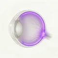
Typical findings of RCH include an orange-red mass with dilated and tortuous feeding and draining vessels. It occurs in the peripheral retina in 85% of cases and near the optic disc in 15%. Pre-treatment visual acuity is maintained at 20/20 or better in 77% of patients.
Major Lesions in VHL Disease
Retinal capillary hemangioma (RCH): Occurs in 50–60% of VHL patients. Characterized by an orange-red tumor and dilated afferent and efferent vessels.
Central nervous system hemangioblastoma: Occurs in the cerebellum, medulla oblongata, and spinal cord. Causes headache and ataxia.
Renal cell carcinoma (RCC): VHL-associated RCC is often bilateral and multifocal. Affects life prognosis.
Pancreatic neuroendocrine tumors and cysts: Occur multifocally throughout the pancreas and may cause exocrine pancreatic insufficiency.
Stages and Progression of RCH
Early stage (small tumor): Small orange-red punctate mass. Mild dilation of afferent and efferent vessels. No subjective symptoms.
Intermediate stage: Tumor enlargement, appearance of exudate and hard exudates. Visual acuity decreases when macular edema occurs.
Advanced stage (large tumor, exudative RD): Complicated by exudative retinal detachment. Extensive exudation and marked decrease in visual acuity.
Ultra-widefield fluorescein angiography (FA) is useful for detecting peripheral RCH, characterized by early hyperfluorescence and late leakage2). Since a single patient may have up to 11 tumors, ultra-widefield imaging that allows visualization of the entire retina is important for diagnosis.
QWhy is it difficult to treat juxtapapillary retinal capillary hemangioma?
A
Because juxtapapillary RCH is adjacent to the optic disc, laser or cryotherapy carries a high risk of optic nerve damage and visual field defects. In addition, exudation tends to spread to the macula, and vision loss may persist after treatment. For details, see the section on “Standard Treatment”.
The direct cause of RCH is a loss-of-function mutation in the VHL gene. The VHL gene is located on the short arm of chromosome 3 (3p25-26) and encodes pVHL (VHL protein), which consists of 232 amino acids 1).
Inheritance pattern and mutations: VHL disease is inherited in an autosomal dominant manner. Tumorigenesis occurs when both a germline mutation in the VHL gene and a somatic mutation in tumor cells (two-hit model) are present. The type of mutation (missense, nonsense, deletion, insertion) influences the clinical phenotype and is reflected in the Type classification 1).
Dilated fundus examination: A detailed examination including the entire peripheral retina. In VHL disease, it is important to confirm bilateral and multiple lesions. Up to 11 tumors may be present in one eye.
Systemic evaluation of VHL disease requires multi-organ imaging, and a systematic surveillance protocol is recommended1).
Examination Item
Target Lesion
Recommended Starting Age
MRI (head and spine)
CNS hemangioblastoma
From age 11
Abdominal MRI/CT
RCC and pancreatic tumors
From age 16
Dilated fundus examination
RCH
From 1 year old
Catecholamine measurement
Pheochromocytoma
From 5 years old
Genetic testing: Confirm VHL gene mutations using Southern blotting, FISH, MLPA, etc. 1). Genetic testing is strongly recommended when VHL disease is clinically suspected.
68Ga-DOTATOC PET-CT: Useful for whole-body assessment using somatostatin receptor (SSTR) expression 3). VHL-associated tumors highly express SSTR (SSTR4: 100%, SSTR1/2/5: 89%) 3), and this method is gaining attention as functional imaging.
Bhende P et al. (2022) performed vitrectomy, tumor resection, and silicone oil (SO) tamponade for tractional and exudative retinal detachment associated with bilateral RCH in a 40-year-old male 4). Postoperative corrected visual acuity was 6/36. Combined TTT improved FFA findings.
QWhat happens if retinal capillary hemangioma is left untreated?
A
If left untreated, the tumor enlarges, leading to accumulation of exudate, formation of hard exudates, and progression to exudative retinal detachment. Further progression may result in neovascular glaucoma, pain, elevated intraocular pressure, and phthisis bulbi5). Early small tumors are more amenable to curative laser treatment, so early detection through regular examinations greatly influences visual prognosis.
pVHL (232 amino acids) functions as a subunit of the E3 ubiquitin ligase complex consisting of elongin B, elongin C, and CUL2, and induces ubiquitination and proteasomal degradation of hypoxia-inducible factor α (HIF-α)1).
A well-established pathway is: VHL mutation → loss of pVHL function → impaired ubiquitination of HIF-α → nuclear accumulation of HIF-α → overexpression of angiogenic factors such as VEGF and PDGF → RCH formation1). RCH exhibits a “pseudohypoxic state” in which HIF is constantly activated regardless of oxygen partial pressure.
The true tumor component of RCH is not endothelial cells but stromal cells. These stromal cells with VHL mutations produce VEGF in a HIF-α-dependent manner, driving angiogenesis. Tumor blood vessels are formed by the paracrine action of stromal cells.
VHL-associated hemangioblastomas highly express SSTR. SSTR4 is expressed in 100% of tumors, and SSTR1/2/5 in 89% 3). This receptor expression is mediated by a hypoxia-independent activation pathway of HIF 3). This characteristic makes them a target for diagnosis with 68Ga-DOTATOC PET-CT and for somatostatin analog therapy.
7. Latest Research and Future Perspectives (Investigational Reports)
Belzutifan is an oral molecular targeted drug that selectively inhibits HIF-2α. It was approved by the US FDA in 2021 for VHL disease-associated tumors (RCC, pancreatic neuroendocrine tumors, and RCH) 1). The standard dose is 120 mg orally once daily 2).
The overall response rate (ORR) for VHL disease-associated RCH is reported to be 100% 1, 2), positioning it as a groundbreaking first systemic drug therapy for existing local ocular treatments.
Ercanbrack CW et al. (2024) reported three cases treated with belzutifan 2). In case 1, complete fibrosis of the tumor was confirmed after 21 months of treatment. In case 3, the tumor area decreased from 10.3 mm² to 5.5 mm² over 7 months 2). Monitoring with ultra-widefield FA was useful for quantitative assessment of tumor shrinkage.
The side effect profile is as follows 2).
Anemia: Occurs in 90%. The most frequent side effect.
Fatigue: Observed in 66%.
Discontinuation: Required in about one-third of cases due to side effects 2).
As a stepwise treatment strategy, combination therapy aiming for cure with laser photocoagulation after tumor shrinkage with belzutifan is being investigated2).
Therapeutic effects of lanreotide 120 mg subcutaneously every 28 days have been reported.
Brabo EP et al. (2024) reported a marked decrease in SUVmax from 15.6 to 4.8 on 68Ga-DOTATOC PET-CT in VHL patients treated with lanreotide3). An average of 10.4 hemangioblastomas per patient were present, suggesting the potential of lanreotide to suppress tumor growth3).
Somatostatin analogs act on hemangioblastomas through a pathway different from HIF-2α inhibitors, suggesting the possibility of future combination therapy3).
This is a functional imaging diagnosis utilizing SSTR expression, and is expected to depict small lesions that are difficult to detect with conventional MRI/CT 3). Studies are underway to verify its usefulness as a systemic surveillance tool for VHL disease.
Bajaj S, Gandhi D, Nayar D, Serhal A. Von Hippel-Lindau Disease (VHL): Characteristic Lesions with Classic Imaging Findings. J Kidney Cancer VHL. 2023;10(3):23-31. doi:10.15586/jkcvhl.v10i3.293.
Ercanbrack CW, Elhusseiny AM, Sanders RN, Santos Horta E, Uwaydat SH. Belzutifan-induced regression of retinal capillary hemangioblastoma: A case-series. Am J Ophthalmol Case Rep. 2024;33:102011. doi:10.1016/j.ajoc.2024.102011.
Brabo EP, Altino de Almeida S, Rafful PP, Rosado-de-Castro PH, Vieira L. Expression of somatostatin receptors in hemangioblastomas associated with von Hippel-Lindau disease as a novel diagnostic, therapeutic, and follow-up opportunity: A case report and literature review. Arch Endocrinol Metab. 2024;68:e230181. doi:10.20945/2359-4292-2023-0181.
Bhende P, Kashyap H, Nadig RR. Surgical management of complicated retinal detachment in a case of retinal hemangioblastoma. Indian J Ophthalmol. 2022;70(8):3167. doi:10.4103/ijo.IJO_1161_22.
Naseripour M, Fadakar K, Azimi F, Taherian MM, Naseripour M, Mirshahi R. Retinal Capillary Hemangioblastoma: A Comprehensive Review on Treatments. Ocul Oncol Pathol. 2026;12(1):53-62. doi:10.1159/000550011.
Copy the article text and paste it into your preferred AI assistant.
Article copied to clipboard
Open an AI assistant below and paste the copied text into the chat box.