
Epicanthal fold (Mongolian fold)
1. What is epicanthus (Mongolian fold)?
Section titled “1. What is epicanthus (Mongolian fold)?”Epicanthus is a condition in which the inner corner of the eye is covered by a crescent-shaped fold of skin and orbicularis muscle from the upper eyelid. In East Asians, it is not necessarily abnormal; during adolescence, as the nasal bone grows, it mostly disappears, and only 2 to 3% remain. It is classified as one of the congenital eyelid anomalies, but in Asian infants it is widely recognized as a physiological variation.
It is classified into two types according to the direction of the fold at the inner eye corner (Verdin H, et al. GeneReviews. 2022).
| Type | Fold direction | Main context |
|---|---|---|
| epicanthus (true epicanthal fold) | from outer upper to inner lower | a physiological variation common in East Asians |
| epicanthus inversus (reverse epicanthal fold) | from outer lower to inner upper | often associated with blepharophimosis syndrome and ptosis |
Epicanthus inversus is often formed when ptosis is present together with epicanthus. In East Asians, it is important to distinguish it from blepharophimosis syndrome.
Blepharophimosis syndrome is an autosomal dominant genetic disorder. In addition to a narrowed palpebral fissure, it shows four signs: ptosis, epicanthus inversus, telecanthus, and lateral displacement of the lacrimal puncta. Its basic pathology is congenital hypoplasia of the eyelid tissues and ptosis, and poor development of the nasal bone makes epicanthal folds more likely.
Epicanthus (Mongolian fold) is a physiological variation common in East Asians and is not necessarily abnormal. Most disappear naturally as the nasal bone grows during puberty, and the residual rate is about 2-3%. However, epicanthus inversus associated with blepharophimosis syndrome carries a risk of amblyopia and requires specialist evaluation and management.
2. Main symptoms and clinical findings
Section titled “2. Main symptoms and clinical findings”Findings of true epicanthus (epicanthus)
Section titled “Findings of true epicanthus (epicanthus)”- A crescent-shaped fold covers the inner canthus, running from the superolateral to the inferomedial side (giving the appearance that the inner corner of the eye is hidden)
- The main change is in appearance, and it can be a cosmetic concern
- Because the nasal sclera is covered by the fold, pseudoesotropia may appear
- In the Hirschberg test, the corneal light reflex shows normal alignment, which helps distinguish it from true esotropia
- It is commonly seen in infants and young children before puberty and usually resolves on its own as they grow. In a study of Chinese children, the ratio of inner canthal distance to interpupillary distance (EFDPD) was shown to drop rapidly between ages 7 and 12, then stabilize afterward (Wei N, et al. Aesthetic Plast Surg. 2019; PMID: 30627812)
Findings of epicanthus inversus
Section titled “Findings of epicanthus inversus”- A fold running from the inferolateral to the superomedial side creates a characteristic appearance
- When associated with blepharophimosis syndrome, the following four signs are present
- Epicanthus inversus
- Ptosis (reduced upper visual field, risk of amblyopia)
- Narrowing of the palpebral fissure (shorter horizontal width)
- Increased intercanthal distance (telecanthus)
Associated findings and objective assessment
Section titled “Associated findings and objective assessment”The abnormality at the inner canthus can occur by itself, but it often accompanies ptosis, epicanthus, and palpebral fissure narrowing. The following measurements are used for diagnosis and follow-up.
- Inner canthal distance (ICD): Birth 20±2 mm, age 2 years 26±1.5 mm are considered normal values
- Interpupillary distance (IPD): Birth 39±3 mm, age 2 years 48±2 mm are considered normal values
In cases with blepharophimosis syndrome, superior visual field obstruction due to ptosis can cause amblyopia. When entropion and epiblepharon are present, corneal epithelial damage (superficial punctate keratopathy: SPK) may be seen.
The fold at the inner canthus covers the nasal white of the eye (sclera), so the eyes can appear to be turned inward. This is called pseudoesotropia. With the Hirschberg test (shine a penlight at the center of the cornea and compare the position of the light reflexes between the two eyes), the light reflexes are centered on both corneas, confirming normal eye alignment.
3. Causes and risk factors
Section titled “3. Causes and risk factors”Embryologic factors
Section titled “Embryologic factors”The formation of epicanthus is closely related to underdevelopment of the nasal bone during fetal facial development. After fusion of the frontonasal and maxillary processes, when the nasal bone is low, excess skin on the inner side of the upper eyelid forms a fold that covers the inner canthus. As the nasal bone grows after puberty, the skin is pulled and stretched, and in many cases it resolves on its own.
In people of Asian descent, because a relatively low nasal bridge and flat facial structure persist for a longer period, epicanthal folds are seen in almost all infants and young children.
Genetic and syndromic factors
Section titled “Genetic and syndromic factors”- Blepharophimosis syndrome: A congenital eyelid disorder with autosomal dominant inheritance. A FOXL2 (3q23) gene mutation has been identified as the causative gene, and the basic pathology is congenital hypoplasia of the eyelid tissues and ptosis. Blepharophimosis syndrome caused by FOXL2 mutations includes type I with ovarian insufficiency and type II with normal ovarian function (Méjécase C, et al. Genes. 2021; PMID: 33806295).
- Chromosomal anomaly syndromes: Epicanthal folds are more common in Down syndrome, Turner syndrome, and Williams syndrome.
- Congenital malformations with nasal bone hypoplasia: Congenital malformations in general that are accompanied by underdevelopment of the nasal bone and zygomatic bone are risk factors for epicanthal folds.
Other risk factors
Section titled “Other risk factors”- Prematurity (nasal bone development is immature at birth)
- If the parents have persistent epicanthal folds (familial clustering)
4. Diagnosis and examination methods
Section titled “4. Diagnosis and examination methods”Visual inspection and morphological assessment
Section titled “Visual inspection and morphological assessment”Diagnosis is made mainly by visual inspection. Assess whether a fold is present at the inner canthus, its direction (normal vs reverse type), and its degree (mild, moderate, or severe). At the same time, confirm the horizontal width of the palpebral fissure and record whether the fissure is narrowed.
Objective measurements
Section titled “Objective measurements”- Inner canthal distance (ICD): Measure the distance between both inner canthi with a ruler or calipers. Compare it with age-based normal values (birth: 20±2 mm, age 2: 26±1.5 mm) to determine whether telecanthus is present.
- Interpupillary distance (IPD): Essential for distinguishing telecanthus (increased ICD with normal IPD) from hypertelorism (both ICD and IPD increased). Normal values are 39±3 mm at birth and 48±2 mm at age 2.
- MRD-1/MRD-2: Evaluate whether ptosis is present and how severe it is. If MRD-1 is 3.5 mm or less, manage as ptosis.
- Levator function test: Needed to assess the severity of congenital ptosis. In cases with blepharophimosis syndrome, levator function is often reduced.
Evaluation of associated lesions
Section titled “Evaluation of associated lesions”- Slit-lamp microscopy: Check for corneal epithelial damage (SPK) when entropion or epiblepharon is present.
- Refraction testing and vision testing: In cases with blepharophimosis syndrome, amblyopia screening is essential. If ptosis is present, perform vision testing and assess for anisometropia and deprivation amblyopia.
- Family history: Because blepharophimosis syndrome is inherited in an autosomal dominant pattern, check the eyelid shape of the parents and siblings.
Differential diagnosis
Section titled “Differential diagnosis”| Differential diagnoses | Key findings | ICD | IPD |
|---|---|---|---|
| physiological epicanthus | crescent-shaped fold, disappears with growth | normal to slightly enlarged | normal |
| blepharophimosis syndrome | the four signs of reverse epicanthus + ptosis + narrowed palpebral fissures + telecanthus | enlarged | normal |
| telecanthus | increased inner canthal distance and elongated medial canthal ligament, with normal interpupillary distance | enlarged | normal |
| Hypertelorism | With widened interorbital distance | Widened | Widened |
| Infantile epiblepharon | Contact between the eyelashes and the cornea due to excess lower eyelid skin | Normal | Normal |
Blepharophimosis syndrome is an autosomal dominant genetic disorder with four features: epicanthus inversus, ptosis, blepharophimosis (shortened horizontal palpebral fissure), and telecanthus. FOXL2 gene mutations are known to cause it, and the underlying problem is congenital hypoplasia of the eyelid tissues. Because ptosis can cause visual impairment (amblyopia), early ophthalmologic evaluation and consideration of surgery are important.
5. Standard treatment
Section titled “5. Standard treatment”Conservative management (observation)
Section titled “Conservative management (observation)”For physiologic epicanthus, observation until puberty is the basic approach. It often resolves naturally as the nasal bones grow, and the residual rate is only about 2–3%.
If pseudostrabismus is suspected, give the parents enough explanation (how it differs from true esotropia) and perform regular strabismus evaluations. Follow-up every 6 months to 1 year is desirable to check whether esotropia has developed.
Surgical treatment
Section titled “Surgical treatment”Treatment is limited to surgical correction; drug therapy is not indicated.
Indications for surgery
Section titled “Indications for surgery”- If epicanthus remains after puberty and cosmetic improvement is desired
- Reverse epicanthus associated with blepharophimosis syndrome (performed at the same time as ptosis surgery or in stages)
- Cases of ptosis with a risk of amblyopia (consider early intervention at 3–5 years of age)
Main procedures for epicanthoplasty
Section titled “Main procedures for epicanthoplasty”- Y-V plasty: The most basic procedure. The fold is released by making a Y-shaped incision and closing it in a V shape. It is simple and the scar is relatively inconspicuous.
- Z plasty: Two skin flaps are exchanged through a Z-shaped incision. It can change the direction of the skin and is also useful for releasing scar contracture. The effect can be adjusted by the angle and direction of the incision.
- Mustardé method: a classic procedure for reverse epicanthus. It is sometimes used together with Y-V plasty. In a case series of blepharophimosis syndrome, Mustardé method double Z-plasty and transnasal intercanthal prolene sutures reduced the postoperative intercanthal distance from 41.2 mm to 31–34 mm, and also increased the horizontal palpebral fissure width (Mandal SK, et al. J Clin Diagn Res. 2017; PMID: 28511421).
- Medial canthal tendon plication: shortens the medial canthal tendon to correct the intercanthal distance when telecanthus is present.
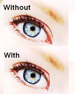
- Skin redraping: a recent mainstream technique for epicanthoplasty in Asians. In a series of 156 cases, it was reported to be minimally invasive, with a scar visibility rate of 0.5% and a recurrence rate of less than 1% (Mo YW, Jung GY. Ann Plast Surg. 2021; PMID: 34559709).
Surgical plan for blepharophimosis syndrome
Section titled “Surgical plan for blepharophimosis syndrome”The medial canthal tendon is plicated, and the soft tissue at the inner corner of the eye is shortened with plastic surgery techniques. If ptosis or entropion is present, surgery for those complications is performed at the same time or in stages.
As for timing, if ptosis with a risk of amblyopia is present, early surgery at age 3 to 5 years is recommended. If the goal is only cosmetic, it is often done around preschool age.
Management of complications
Section titled “Management of complications”- With entropion: if the eyelashes touch the cornea, entropion surgery (Hotz method, etc.) is performed.
- With corneal epithelial damage: for punctate superficial keratopathy (SPK), use artificial tears and corneal epithelium-protective eye drops (such as sodium hyaluronate eye drops).
- With amblyopia: refractive correction and amblyopia training with an eye patch are carried out in parallel.
Physiologic epicanthal fold (Mongolian fold) is generally observed until puberty and often resolves naturally as the nasal bone grows. If it remains and cosmetic improvement is desired, epicanthoplasty is considered after puberty (generally from junior high school age). On the other hand, when reverse epicanthal fold associated with blepharophimosis syndrome carries a risk of amblyopia due to ptosis, early surgical intervention at 3 to 5 years of age is recommended.
6. Pathophysiology and detailed mechanism of onset
Section titled “6. Pathophysiology and detailed mechanism of onset”Embryologic background
Section titled “Embryologic background”During fetal facial development, the frontonasal prominence and maxillary prominences fuse to form the nose, upper lip, and upper eyelid structures. When nasal bone development is insufficient during this process, excess skin on the medial upper eyelid forms a fold that covers the inner canthus. As the nasal bone grows and projects during puberty, the skin is pulled and stretched toward the nasal bridge, and the fold resolves. In East Asians, a relatively low nasal bridge tends to persist, and epicanthal folds are common in infancy.
Relationship between the nasal bone and epicanthus
Section titled “Relationship between the nasal bone and epicanthus”When the nasal root is low, laxity and excess skin develop around the inner canthus. This excess skin folds at the inner canthus to form the fold. With an increase in nasal bridge height (pubertal growth), the skin is pulled, and the fold becomes flatter and disappears.
Pathophysiology of blepharophimosis syndrome
Section titled “Pathophysiology of blepharophimosis syndrome”In blepharophimosis syndrome, congenital hypoplasia of the eyelid tissues (including dysfunction of the levator and frontalis muscles) coexists with underdevelopment of the nasal bone. This causes four findings to appear together: reverse epicanthal fold, ptosis, narrowed palpebral fissures, and telecanthus.
The FOXL2 (forkhead box L2) gene is located at 3q23 and encodes a transcription factor involved in the development of the eyelids and ovaries. Blepharophimosis syndrome caused by FOXL2 mutations is classified into type I, with ovarian dysfunction, and type II, in which ovarian function is preserved. Both familial and sporadic forms occur.
Mechanism of pseudoesotropia
Section titled “Mechanism of pseudoesotropia”When the epicanthal fold covers the nasal sclera (white of the eye), the cornea appears shifted toward the nose even though the eyes are aligned. This is the mechanism of pseudoesotropia. The Hirschberg test or alternate cover test is useful for distinguishing it from true esotropia, and confirms that eye position is straight.
7. Recent research and future prospects
Section titled “7. Recent research and future prospects”In the field of research on epicanthus and blepharophimosis syndrome, the following advances have been reported.
- Advances in FOXL2 mutation analysis: Studies on the genotype-phenotype correlation of FOXL2 in blepharophimosis syndrome have accumulated, and research is progressing on predicting the presence or absence of ovarian dysfunction (type I) from the type of gene mutation.
- Refinements in epicanthoplasty techniques: Through the development of micro-incision approaches and scar-minimizing techniques, procedures that reduce postoperative scarring have been reported. In particular, demand is increasing for better cosmetic results after surgery in procedures performed for aesthetic purposes.
- One-stage repair of blepharophimosis syndrome: Results are still being evaluated for procedures that perform epicanthoplasty and ptosis surgery at the same time (one-stage repair). Comparisons with staged surgery are being made for safety and effectiveness.
- 3D simulation: The potential use of 3D simulation for preoperative surgical planning is being studied, and it may be useful for choosing the appropriate procedure and explaining the expected postoperative outcome to patients.
- Natural history studies of epiblepharon: Large studies from Asia on the spontaneous resolution rate and risk of corneal damage in infantile epiblepharon, which often coexists with epicanthus, have been reported and are helping standardize surgical indications.
Future challenges include integrating genetic diagnosis with phenotype prediction, further refining minimally invasive surgical techniques, and establishing protocols for coordination with amblyopia management.
8. References
Section titled “8. References”- Wei N, Qian X, Bi H, et al. Pseudoesotropia in Chinese Children: A Triphasic Development of the Interepicanthal Folds Distance-to-Interpupillary Distance Ratio and Its Changing Perception. Aesthetic Plast Surg. 2019;43(2):492-499. PMID: 30627812. PubMed
- Méjécase C, Nigam C, Moosajee M, Bladen JC. The Genetic and Clinical Features of FOXL2-Related Blepharophimosis, Ptosis and Epicanthus Inversus Syndrome. Genes (Basel). 2021;12(3):364. PMID: 33806295. PubMed
- Méjécase C, Nigam C, Moosajee M, Bladen JC. The Genetic and Clinical Features of FOXL2-Related Blepharophimosis, Ptosis and Epicanthus Inversus Syndrome. Genes (Basel). 2021 Mar 4;12(3):364. doi:10.3390/genes12030364. PMID:33806295; PMCID:PMC7998575.
- Mandal SK, Mandal A, Fleming JC, Goecks T, Meador A, Fowler BT. Surgical Outcome of Epicanthus and Telecanthus Correction by Double Z-Plasty and Trans-Nasal Fixation with Prolene Suture in Blepharophimosis Syndrome. J Clin Diagn Res. 2017;11(3):NC01-NC04. PMID: 28511421. PubMed
- Mo YW, Jung GY. Surgical Results and Patient Satisfaction After A New Surgical Technique for Asian Medial Epicanthoplasty: A Modified Skin Redraping Method Using a Horizontal Point Incision and Staged ‘Y-Shaped’ Dog Ear Correction. Ann Plast Surg. 2021. PMID: 34559709. PubMed