Anophthalmic Socket Syndrome / Post-enucleation Socket Syndrome is a condition in which the socket becomes atrophic and sunken after enucleation or evisceration. It is characterized by redistribution of orbital fat, ptosis, deep superior sulcus deformity, lower eyelid malposition, and lagophthalmos. In addition to cosmetic problems, it causes difficulty retaining the ocular prosthesis.
The average volume of the eyeball is about 7.2 ml (a sphere 24 mm in diameter), and the total orbital volume is measured at about 24 ml1). The volume deficit after enucleation reaches 7.0–9.0 ml (average 7.9 ml)1). This deficit is replaced with an orbital implant and an ocular prosthesis, but if the replacement is inadequate or if the implant and orbital tissues atrophy over time, socket hollowing occurs.
Post-enucleation socket syndrome (PESS) is defined as a complex set of changes after enucleation, including difficulty retaining the prosthesis and poor cosmetic appearance2).
QWhy does an anophthalmic socket shrink?
A
After enucleation, orbital volume decreases by about 7–9 ml. If the orbital implant is insufficient, or if fat atrophies over time, the orbit becomes sunken and it becomes difficult to retain the prosthesis. After radiation therapy, atrophy tends to progress further, and in children, underdevelopment of the orbital bones is also added.
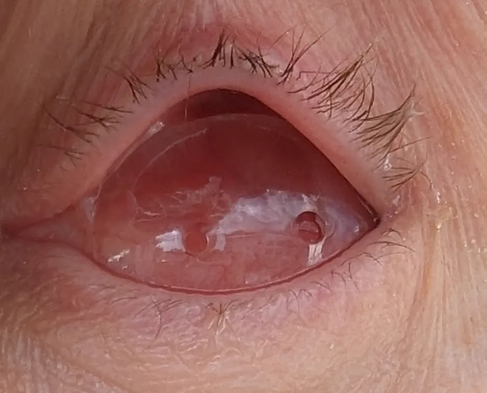
In the left anophthalmic socket 35 days after enucleation, with the upper eyelid lifted, a conformer (a transparent plastic spacer) can be seen in place over the orbital implant. This corresponds to the post-enucleation socket (conformer in place) discussed in section “2. Main symptoms and clinical findings.”
The pathology of anophthalmic socket depression is classified into the following three types.
Type
Main problem
First-line treatment
Conjunctival fornix contracture type
The conjunctival sac is too narrow for the prosthetic eye to fit
Conjunctival fornix expansion surgery (full-thickness skin graft)
Orbital depression type
Insufficient orbital volume
Augmentation procedure (DFG, bone graft, artificial materials)
Mixed type
Combination of both
Stepwise treatment (expansion surgery → augmentation surgery)
Conjunctival socket contracture type
Main condition: The conjunctival socket becomes narrow and shallow, and the prosthetic eye no longer fits properly.
Treatment: Conjunctival socket expansion surgery is indicated, in which the conjunctival socket is dissected upward, downward, left, and right, and a full-thickness skin graft is wrapped around the prosthetic eye and inserted.
Orbital hollowing type
Main condition: Due to insufficient orbital volume, the orbital area moves backward and becomes sunken.
Treatment: Augmentation with autologous tissue (dermal fat, iliac bone, costal cartilage) or artificial materials (hydroxyapatite, silicone) is indicated.
Mixed type
Main condition: Both conjunctival socket contracture and orbital hollowing are present.
Treatment: Conjunctival socket expansion surgery is usually performed first, followed by stepwise orbital augmentation surgery.
Sunken appearance confirmed by inspection and palpation of the orbit
Quantitative assessment with a Hertel exophthalmometer (compared with the healthy side)
After radiotherapy (and after surgery for malignant tumors such as retinoblastoma), scarring of the orbital tissues is added, and the hollowing becomes especially pronounced. In children, facial asymmetry and orbital underdevelopment are also seen.
QWhy does my prosthetic eye fall out so often? What is the cause?
A
It can be caused by contracture of the conjunctival sac (the space that holds the prosthetic eye becomes smaller), a sunken orbit (the lower support is not sufficient), or both. It is important to see an ophthalmologist (an oculoplastic surgeon) so they can assess the size of the conjunctival sac and the orbital volume.
Anophthalmic socket atrophy occurs as a long-term complication after enucleation or evisceration. The rate of onset and the speed of progression are affected by various factors.
Type of orbital implant and risk of exposure: After enucleation using porous polyethylene (MEDPOR) or hydroxyapatite, implant exposure occurs in 1.5–21.6%; after evisceration, it occurs in 0–3.3%3). Implant exposure or movement causes secondary changes in orbital volume and worsens sunken appearance.
Pediatric cases: Normal development of the orbital bones requires the presence of the eye, and enucleation in early childhood can lead to underdevelopment of the orbit. Because orbital volume reaches about 80% of adult size by age 56), management during this period is especially important.
After radiation therapy: Fibrosis and vascular damage in the orbital tissues accelerate atrophy. In children treated during the growth period, underdevelopment of the facial bones is also added.
No implant / implant extrusion: If no orbital implant is inserted, or if the implant is extruded, orbital fat atrophy and redistribution progress and the sunken appearance becomes more noticeable.
Indicated when the conjunctival sac is contracted or narrowed and the prosthetic eye does not fit.
Procedure:
Dissect the conjunctival sac upward, downward, and to the left and right (four directions) to secure enough space
Wrap a full-thickness skin graft taken from the groin or lower abdomen inside out around a thin prosthetic eye and insert it
Place it so the graft and prosthetic eye fit within the conjunctival sac
Fix the inferior fornix of the conjunctival sac firmly and deeply to the periosteum of the inferior orbital rim (if fixation is inadequate, the prosthetic eye is more likely to protrude)
Types and selection of orbital implants (Orbital Implant)
DFG is an orbital reconstruction method using autologous tissue; it does not cause foreign-body reaction and is an excellent procedure with a low risk of orbital implant exposure4).
Graft components:
Dermal button (20–25 mm diameter)
Fat portion (20–35 mm thickness)
Harvest site (hairless area preferred):
Buttock (most common)
Inner thigh
Abdomen
Groin
Set the graft size 10–30% larger than the orbital volume. If it is too large, it can cause pressure necrosis; if too small, it can lead to atrophy and recurrent hollowing.
Key points of the procedure:
Attach the extraocular muscles to the dermal button with horizontal mattress sutures to ensure movement of the prosthesis
After surgery, place a conformer and remove it 3–4 weeks later
After the conjunctival epithelium covers the dermal surface in 4 to 6 weeks, a prosthetic eye is fitted
DFG outcomes5):
Indicator
Primary DFG
Secondary DFG
Good eyelid position
83.3%
37.5%
Fat atrophy rate
5–10%
20–40%
Good prosthetic eye movement
83.3%
100%
Complication rate (overall)
58.8% (most were mild and resolved on their own)
—
Primary DFG (performed at the same time as enucleation) has a better prognosis for eyelid position than Secondary DFG (secondary reconstruction)5).
QWhat kind of surgery is dermis fat grafting?
A
This is an autologous tissue graft in which dermis and fat are taken from the buttock or inner thigh and transplanted into the orbit. There is no foreign-body reaction, and the risk of implant exposure is low. The conjunctival epithelium covers the dermal surface in 4–6 weeks, after which the prosthetic eye is fitted. If it shrinks again, regrafting is also possible.
Comparison of materials for correcting socket hollowing (augmentation)
Autologous tissue, low exposure risk, possible regrafting
Risk of atrophy over time
Iliac bone block
Fits bone atrophy and is firm
Partial resorption and donor-site complications
Costal cartilage
Autologous tissue and easy to shape
Leaves a scar on the chest
Hydroxyapatite
Osteoconductive and stable
Risk of foreign body reaction and surface exposure
Silicone block
Inexpensive and easy to process
Risk of movement and foreign body reaction
Microsurgical flap
Can transplant large amounts of tissue
Highly invasive, long operation
For orbital bony atrophy, grafting an iliac bone block or crushed iliac bone is suitable. For less severe depression, a dermis-fat graft is relatively easy to harvest and also makes the prosthetic socket softer, so it is a good option.
Selection of donor site: Even if scars remain on the iliac crest or groin, the skin incision can be placed where it can be hidden by underwear or a swimsuit.
Conjunctival sac expansion surgery
Indications: Contracted conjunctival sac type.
Procedure: Wrap a full-thickness skin graft inside out around a thin ocular prosthesis and insert it. Fix the lower fornix deeply to the periosteum of the infraorbital rim.
Donor site: Groin or lower abdomen (a position hidden by underwear or a swimsuit).
Dermis-fat graft (DFG)
Indications: Moderate orbital hollowing, salvage for exposed orbital implant, chronic pain.
Procedure: Attach the extraocular muscles to the dermal button. Fit the prosthesis after leaving the conformer in place for 3–4 weeks.
Advantages: Can be re-implanted if atrophy recurs. Low risk of exposure.
Bone grafts and artificial materials
Indication: Severe socket depression with marked bony atrophy.
Material selection: Iliac bone (bony atrophy), hydroxyapatite (stability), silicone block (low cost; deep insertion is important).
Note: If silicone is not inserted deeply, there is a risk of movement and exposure.
QCan corrective surgery for the ocular socket be done more than once?
A
Dermis-fat grafting can be repeated if atrophy recurs. However, repeated surgery may lead to scarring of the conjunctival sac. The choice of material should be discussed with an oculoplastic specialist.
6. Pathophysiology and detailed mechanism of onset
The volume loss after eye removal reaches 7.0–9.0 mL (average 7.9 mL)1). Inserting an orbital implant makes up much of this volume, but the implant alone cannot fill the entire orbit, so the remaining deficit is covered by an artificial eye (an external prosthesis placed over the socket base). If the implant is inadequate, extruded, or displaced, or if the orbital fat shrinks over time, a depression develops.
Radiation to the orbit causes fibrosis and vascular damage in orbital tissues. Fibrotic tissue loses elasticity and shrinks, causing the entire socket to become smaller. In children, radiation can also impair orbital bone growth, leading to marked facial asymmetry later in adulthood.
Normal development of the orbital bones requires the presence of the eye. When the eye is lost, the mechanical stimulus to the orbit is lost and orbital bone growth is delayed. By age 5, orbital volume reaches about 80% of the adult size6), so during this period it is especially important to manage the orbital implant and artificial eye to maintain orbital volume. As the child grows, the size of the artificial eye and implant must be adjusted regularly.
Exposure occurs when the conjunctiva and Tenon’s capsule over the implant become thin and necrotic. Porous materials promote tissue integration through vessel ingrowth, but exposure risk remains if the surface tissues are thin or if blood flow is impaired by surgical manipulation. The dermis-fat graft has the lowest exposure risk because blood flow is maintained in autologous tissue4).
Fat atrophy begins immediately after removal of the eye, and without an implant the hollowing progresses especially rapidly. Even with an implant, hollowing gradually advances because of aging, gravity, and pressure from the weight of the artificial eye. This change is further accelerated after radiation therapy and during periods of growth.
Long-term outcomes of DFG: In a case series of 34 cases (Jovanovic et al.), a complication rate of 58.8% was reported, but most were mild and resolved on their own4). DFG salvage for implant exposure was indicated in 67.7% of cases, making it an effective salvage option4).
5-fluorouracil (5-FU) injections: An approach has been reported in which an antimetabolite is used as a pre-treatment for severe contracted socket, softening scar tissue before DFG is performed7).
Use for chronic pain: There are reports that chronic socket pain improves by removing the orbital implant and replacing it with DFG8).
Expandable hydrogel implant (HEMA): Research is being conducted on stepwise expanding HEMA implants to promote orbital growth in children.
Future challenges: It is necessary to identify predictors of long-term atrophy in DFG, standardize the optimal graft size, and compare long-term outcomes by material of the ocular prosthesis base (RCT).
Schmitzer S, Simionescu C, Alexandrescu C, Burcea M. The Anophthalmic Socket - Reconstruction Options. Journal of medicine and life. 2014;7 Spec No. 4(Spec Iss 4):23-9. PMID:27478515; PMCID:PMC4962761.
Aggarwal H, Singh K, Kumar P, Alvi HA. A multidisciplinary approach for management of postenucleation socket syndrome with dermis-fat graft and ocular prosthesis: a clinical report. Journal of prosthodontics : official journal of the American College of Prosthodontists. 2013;22(8):657-60. doi:10.1111/jopr.12051. PMID:23552097.
Custer PL, Kennedy RH, Woog JJ, Kaltreider SA, Meyer DR. Orbital implants in enucleation surgery: a report by the American Academy of Ophthalmology. Ophthalmology. 2003;110(10):2054-2061. doi:10.1016/s0161-6420(03)00857-1. PMID:14522788.
Jovanovic N, Carniciu AL, Russell WW, Jarocki A, Kahana A. Reconstruction of the orbit and anophthalmic socket using the dermis fat graft: a major review. Ophthalmic Plast Reconstr Surg. doi:10.1097/iop.0000000000001610.
Nentwich MM, Schebitz-Walter K, Hirneiss C, Hintschich C. Dermis fat grafts as primary and secondary orbital implants. Orbit (Amsterdam, Netherlands). 2014;33(1):33-8. doi:10.3109/01676830.2013.844172. PMID:24195744.
Bentley RP, Sgouros S, Natarajan K, Dover MS, Hockley AD. Normal changes in orbital volume during childhood. Journal of neurosurgery. 2002;96(4):742-6. doi:10.3171/jns.2002.96.4.0742. PMID:11990816.
Priel A, Oh SR, Whipple KM, et al. Use of antimetabolites in the reconstruction of severe anophthalmic socket contraction. Ophthalmic Plast Reconstr Surg. 2012;28:409-412.
Shams PN, Bohman E, Baker MS, Maltry AC, Kopp ED, Allen RC. Chronic anophthalmic socket pain treated by implant removal and dermis fat graft. The British journal of ophthalmology. 2015;99(12):1692-6. doi:10.1136/bjophthalmol-2014-306585. PMID:26041123.
Copy the article text and paste it into your preferred AI assistant.
Article copied to clipboard
Open an AI assistant below and paste the copied text into the chat box.
