Pediatric uveitis is a relatively rare disease, with an estimated prevalence of about 30 per 100,000 population2). Non-infectious uveitis accounts for 69–95% of pediatric uveitis2). The most common identifiable cause is juvenile idiopathic arthritis (JIA), accounting for 41–47% of all cases2).
Glaucoma is a serious complication of pediatric uveitis. 5–42% of pediatric uveitis patients develop glaucoma or ocular hypertension, and 5.3–19% of all pediatric glaucoma cases are due to uveitis. According to the World Glaucoma Association (WGA) classification, secondary glaucoma due to uveitis is classified as “secondary glaucoma due to acquired factors”1).
Ocular complications including cataract, glaucoma, and macular edema have been reported in up to 76% of pediatric uveitis cases2). In a study of 166 cases by Cann et al., children with JIA-associated uveitis (JIA-U) developed glaucoma significantly more often than those with idiopathic uveitis (p=0.002)2).
QHow are juvenile idiopathic arthritis and secondary glaucoma due to uveitis related?
A
Juvenile idiopathic arthritis is a chronic arthritis of unknown cause that begins before age 16, and it is the most common cause of pediatric uveitis. Uveitis develops in about 20% of oligoarticular JIA and about 5% of polyarticular JIA. In JIA-U, chronic anterior uveitis causes trabecular meshwork damage and iris synechiae, and secondary glaucoma complicates 19–25% of cases3). The incidence of glaucoma is significantly higher in JIA-U compared to idiopathic uveitis2).
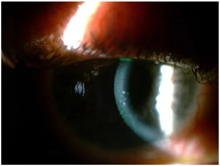
Ioannis Halkiadakis; Kalliroi Konstantopoulou; Vasilios Tzimis; et al. Update on Diagnosis and Treatment of Uveitic Glaucoma. Journal of Clinical Medicine. 2024 Feb 20. Figure 1. PMCID: PMC10931771. License: CC BY.
Slit-lamp photograph showing anterior segment findings with keratic precipitates and anterior chamber inflammation. The deposits on the corneal endothelium and anterior chamber inflammation indicate activity of uveitis-associated glaucoma.
Juvenile idiopathic arthritis-associated uveitis is described as “white uveitis.” It is often accompanied by little ciliary injection or eye pain, and visual loss is also difficult to notice. Therefore, detection tends to be delayed. Ophthalmology consultation often occurs around age 10, but the actual onset of uveitis is estimated to be between 1 and 6 years of age.
Band keratopathy: Often already present at the initial examination
Complicated cataract: The most common ocular complication, with an incidence of 0.05 per eye-year (EY) 2)
Posterior synechiae: The most common complication at initial examination, seen in 18.4% of cases 2)
Keratic precipitates: Typically fine, non-granulomatous deposits
Intraocular Pressure-Related Findings
Elevated intraocular pressure: In juvenile idiopathic arthritis-associated uveitis, elevated intraocular pressure is significantly more common than in idiopathic uveitis (p=0.05) 2)
Glaucoma complication: The rate of glaucoma in juvenile idiopathic arthritis-associated uveitis varies from 9.4% to 30% depending on the report 2)
Optic disc cupping: Glaucomatous changes often appear more than 10 years after the onset of uveitis, but can also progress rapidly.
Juvenile idiopathic arthritis is the most important underlying disease. Juvenile idiopathic arthritis is clinically classified into systemic and articular types, and the articular type is further classified into oligoarticular and polyarticular types. Uveitis most commonly occurs in the oligoarticular type, affecting about 20% of patients. Antinuclear antibody (ANA) is positive in approximately 80% of juvenile idiopathic arthritis cases with uveitis, and has high diagnostic value.
Secondary glaucoma occurs in 19–25% of patients and is one of the poor prognostic factors 3). The mechanisms of glaucoma development are broadly divided into the following three categories.
Acute glaucoma attack due to circumferential posterior synechiae of the iris
Formation of anterior synechiae in the angle due to chronic iridocyclitis
Steroid-induced intraocular pressure elevation occurs in about 20% of children receiving corticosteroid therapy. If left untreated, steroid-induced ocular hypertension tends to progress to glaucoma more rapidly in children than in adults.
QHow do we differentiate between steroid-induced ocular hypertension and uveitis-induced ocular hypertension?
A
Elevated intraocular pressure in the presence of active inflammation is usually secondary to uveitis. In such cases, increasing the frequency of steroid administration to suppress inflammation will lower the intraocular pressure. On the other hand, if intraocular pressure is high despite controlled inflammation, it is likely a steroid response, requiring reduction or discontinuation of steroids.
The diagnosis of childhood glaucoma uses the WGA diagnostic criteria 1). It is defined as meeting two or more of the following five items: intraocular pressure >21 mmHg, progressive increase in cup-to-disc ratio, corneal findings (Haab striae, increased corneal diameter), abnormal elongation of axial length, and glaucomatous visual field defects 1).
The following points should be noted during examination:
Gonioscopy: Differentiation between open-angle and closed-angle is directly linked to treatment strategy.
Assessment of anterior chamber inflammation: Evaluate the degree of anterior chamber cells and flare.
Differentiation between inflammatory and steroid-induced ocular hypertension.
In infants and young children, examination under general anesthesia is often required 1). In patients with juvenile idiopathic arthritis-U, those referred to tertiary care centers had a significantly higher likelihood of developing visual impairment >0.3 logMAR (p=0.03), and posterior synechiae at initial visit was a risk factor for visual impairment ≥1.0 logMAR (p=0.01) 2).
Treatment of systemic inflammatory disease and control of uveitis form the basis for prevention and progression suppression of secondary glaucoma. Collaborative management with rheumatology is essential. In pediatric uveitic glaucoma, the success rate with eye drops alone is low, at 17–26%.
In children, even with eye drops, attention must be paid to the dose relative to body weight and body surface area 1). Punctal occlusion and use of gel formulations are useful for reducing side effects.
First-Line Drugs
Beta-blockers (timolol): First-line monotherapy. Gel-forming solutions administered once daily improve adherence. Contraindicated in children with asthma, arrhythmia, or apnea.
Carbonic anhydrase inhibitors (dorzolamide): Most commonly used in combination with beta-blockers. Oral acetazolamide is also effective but has many side effects.
Other Medications
Prostaglandin analogs: Not associated with increased risk of uveitis or cystoid macular edema. Administered once daily.
Alpha-2 agonists (brimonidine): Absolutely contraindicated in children under 2 years. Relatively contraindicated in children under 6 years or weighing less than 20 kg1)4).
Miotics (pilocarpine): Contraindicated in active uveitis.
If medical treatment is insufficient, surgical treatment is considered1). Angle surgery is the first choice; if unsuccessful, filtering surgery or GDD may be considered1)4).
Goniotomy improves aqueous outflow by incising the trabecular meshwork from the inside. In a study of 40 eyes with pediatric uveitic glaucoma, surgical success was achieved in 72% at final follow-up. Mean intraocular pressure in the success group decreased from 35.8 to 14.7 mmHg. Phakic eyes and age under 10 years were predictors of success.
Trabeculotomy reduced mean postoperative intraocular pressure from 31.4 to 15.0 mmHg, and the number of glaucoma medications decreased from 4.2 to 0.4.
Filtering surgery typically uses mitomycin C (MMC) as an adjunct. Notably, the combination of TNF inhibitors improved outcomes: the 5-year success rate with MMC alone was only 16%, whereas with MMC plus TNF inhibitors it was 73%.
Glaucoma drainage device (GDD) surgery: in a study of 27 eyes with Molteno GDD, 90% achieved success (IOP 6–22 mmHg). With the Ahmed glaucoma valve, IOP was maintained at 7–18 mmHg in all eyes.
Cyclodestructive procedures are considered when the above treatments are ineffective, but the success rate is low at 32%, and there is a risk of exacerbating inflammation1).
QWhat are the surgical outcomes for pediatric uveitic glaucoma?
A
Outcomes vary greatly by procedure. Goniotomy has a reported success rate of 72% (at 98.9 months), and trabeculotomy 81.8%. For filtering surgery, the 5-year success rate with MMC alone is low at 16%, but improves to 73% with TNF inhibitors. GDD surgery with Molteno GDD has a 90% success rate. Cyclodestructive procedures have the lowest success rate at 32%. Overall, outcomes tend to be worse than in adult uveitic glaucoma.
Trabecular meshwork (TM) obstruction is the most common mechanism3). Breakdown of the blood-aqueous barrier (BAB) allows inflammatory cells to enter the aqueous humor, where they become trapped in the outflow pathways along with normal serum components3). Additionally, swelling of the trabecular beams and deposition of inflammatory debris reduce TM permeability. Trabeculitis directly impairs TM function.
Chronic inflammation can lead to permanent scarring of the trabecular meshwork, resulting in irreversible outflow obstruction. Glaucoma occurs in approximately 20% of uveitis patients3).
The angle is mechanically obstructed by formation of peripheral anterior synechiae (PAS), pupillary block due to posterior synechiae, or formation of fibrous membranes. Circumferential posterior synechiae can cause acute glaucoma attacks.
Long-term steroid administration alters the extracellular matrix of the trabecular meshwork (TM), increasing resistance to aqueous humor outflow. In children, if steroid-induced ocular hypertension is left untreated, glaucoma tends to progress earlier and more rapidly.
The introduction of biologic agents has improved visual prognosis in pediatric uveitis2). In a study by Cann et al., the use rate of biologics was 34.9%, showing improvement in visual impairment rates and ocular complication rates compared to previous reports 2). Adalimumab was the most commonly used biologic, administered to 31.3% of patients 2).
The impact of TNF inhibitors on surgical outcomes for uveitic glaucoma is noteworthy. The 5-year success rate of filtration surgery combining MMC and TNF inhibitors was 73%, significantly better than the 16% with MMC alone.
The application of minimally invasive glaucoma surgery (MIGS) in children is also advancing. Promising results of gonioscopy-assisted transluminal trabeculotomy (GATT) for juvenile idiopathic arthritis-associated glaucoma have been reported, with intraocular pressure reductions of 40–66% in three eyes.
Future challenges include conducting RCTs specific to pediatric uveitic glaucoma, evaluating the long-term effects of biologics on intraocular pressure, and accumulating safety and efficacy data for MIGS in children.
Cann M, Ramanan AV, Crawford A, Dick AD, Clarke SLN, Rashed F, et al. Outcomes of non-infectious Paediatric uveitis in the era of biologic therapy. Pediatr Rheumatol Online J. 2018;16(1):51. doi:10.1186/s12969-018-0266-5. PMID:30081917. PMCID:PMC6080499.
Bodh SA, Kumar V, Raina UK, et al. Inflammatory glaucoma. Oman J Ophthalmol. 2011;4(1):3-9.
Pazos M, Traverso CE, Viswanathan A; European Glaucoma Society. European Glaucoma Society - Terminology and guidelines for glaucoma, 6th Edition. Br J Ophthalmol. 2025;109(Suppl 1):1-212. doi:10.1136/bjophthalmol-2025-egsguidelines. PMID:41026937.
Copy the article text and paste it into your preferred AI assistant.
Article copied to clipboard
Open an AI assistant below and paste the copied text into the chat box.