Tube shunt exposure is a complication after glaucoma drainage device (GDD) surgery in which the conjunctiva or patch graft covering the device erodes, exposing the tube or plate to the outside. Implant exposure is a major risk of glaucoma drainage devices and carries a risk of endophthalmitis1).
Glaucoma drainage devices are increasingly used as an alternative filtration surgery to trabeculectomy. In Japan, two types are covered by insurance: the Baerveldt glaucoma implant and the Ahmed glaucoma valve.
Primary Open-Angle Glaucoma PPP: TVT study 5%, ABC study 1–2.9%, AVB study 2–4% 3).
In terms of intraocular pressure control, there is no significant difference between trabeculectomy and tube shunt surgery, but implant exposure and corneal endothelial damage are more common with tube shunt surgery 4). On the other hand, bleb leakage, bleb infection, and endophthalmitis are more common with trabeculectomy4).
QHow often does tube shunt exposure occur?
A
Large studies report an incidence of about 2–5%. In the TVT study, it was 4.7% at 5 years, and a meta-analysis of 3255 eyes reported 2.0±2.6%. Early studies that did not use patch grafts reported rates as high as 30%.
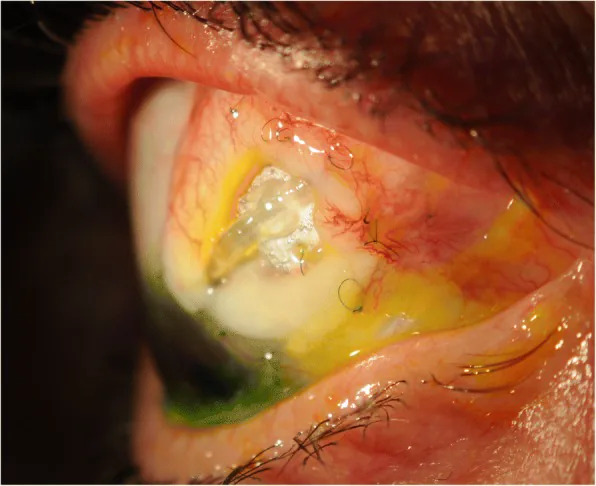
Gurjeet Jutley; Elizabeth Yang; Phillip Bloom. Surgical management of raised intra-ocular tension in the hostile ocular surface - recurrent tube erosion in a patient with systemic sclerosis: a case report. BMC Ophthalmol. 2018 Sep 14;18(Suppl 1):222. Figure 2. PMCID: PMC6157120. License: CC BY.
Clinical photograph showing a tube extender exposed at the site of conjunctival erosion. The clinical appearance of tube shunt exposure is visible, indicating a condition with high infection risk.
Visibility of the tube under the conjunctiva: a condition where the tube is visible under the conjunctiva requires attention; even without exposure, the risk of infection is high
Positive fluorescein staining (Seidel sign): confirms fluid leakage. Used to differentiate tissue thinning from actual exposure.
Patch graft thinning: The patch gradually thins over time, leading to exposure.
Chronic inflammation/ischemia: Uveitic glaucoma and long-term steroid use increase the risk of exposure. In the uveitis group, tube exposure leading to device removal has been reported in 13.3% of cases2).
Prior ocular surgery: A history of trabeculectomy with antifibrotic agents is a risk factor. Sequential glaucoma drainage devices also increase the exposure rate.
Pediatric patients: Frequent eye rubbing, small orbital volume, and increased device mobility are risk factors.
Other: Data on sex and race are inconsistent across studies, likely due to small sample sizes.
Surgical Factors
Placement location: The inferior quadrant has a higher exposure risk than the superior quadrant (12.8% vs 5.4%). The inferonasal quadrant carries the highest risk. Infection risk is also higher in the inferior quadrant.
Patch graft material: The difference in exposure risk depending on the material is under debate. Some reports indicate a reduced risk with double-layer pericardium, while others report an increased risk with bovine pericardium.
Location of conjunctival incision: If the incision line is over the plate, the probability of plate exposure increases.
Device type and size: The material, type, and size of the shunt are not associated with exposure risk.
To prevent tube exposure, it is necessary to cover the tube with patch materials such as preserved sclera or preserved cornea, or with an autologous scleral flap (recommendation grade 1A) 4). Careful conjunctival and scleral suturing is important for preventing exposure.
QShould inferior tube placement be avoided?
A
Basically, the superotemporal quadrant is the first choice. The exposure rate in the inferior quadrant is 12.8%, approximately 2.4 times that in the superior quadrant (5.4%), and the infection risk is also higher. If the superior quadrant is unavailable due to previous surgical wounds, the nasal or inferior quadrant may be considered, but it is recommended to avoid the inferior quadrant as much as possible.
Since late exposure can occur several years after surgery, long-term regular examinations are necessary after glaucoma drainage device surgery. During examinations, carefully observe the device and surrounding tissues.
Eyelid eversion test: Ask the patient to look downward (or upward for inferiorly placed devices) and examine the tube, plate, and covering tissues.
Fluorescein staining: Apply stain over the device and check for a Seidel sign. Differentiate between tissue thinning alone and actual exposure.
Anterior chamber/vitreous evaluation: Check for signs of infection (hypopyon, vitritis)
Covering with patch material is essential to prevent tube exposure4).
Covering method
Characteristics
Notes
Preserved scleral patch
Most common
Thinning over time
Autoscleral flap
No additional material needed
Tube insertion under the flap
Scleral tunnel
No patch needed
Long tunnel is effective
In Baerveldt glaucoma implants, the tube is covered with preserved sclera to prevent postoperative tube exposure. A method of creating an autoscleral flap and inserting the tube has also been reported.
Secure insertion and fixation of both ends of the plate under the rectus muscles is important to prevent plate dislocation. Since the Hoffman elbow is thick and poses a risk of exposure, using a straight tube type is also an option.
The repair techniques for when exposure occurs are as follows.
Tube repositioning: Changing the intraocular insertion site. Repositioning posteriorly (from anterior chamber to ciliary sulcus) shortens the extraocular tube length and reduces the risk of re-exposure.
Tube rerouting: Changing the route from the plate to the intraocular space to alter mechanical forces.
Scleral tunnel creation: Reduces tube mobility and friction with covering tissue. May be difficult in cases of scleral thinning or high myopia.
Secondary patch graft: Reduces the risk of re-exposure by about half compared to direct conjunctival closure alone.
Nylon sutures are recommended. Polyester sutures (such as Mersilene) have a higher re-exposure rate due to increased friction and immune reaction.
QIs there a possibility of re-exposure after tube exposure repair?
A
The re-exposure rate after repair is high, with multiple studies reporting 41–45%. One study reported that 43% required additional surgical intervention after initial repair. The use of a secondary patch graft and choice of suture material (nylon suture recommended) may contribute to reducing the risk of re-exposure.
The pathophysiology of tube exposure differs depending on the timing of onset.
Early Exposure
Onset: Within a few months after surgery
Mechanism: Caused by wound dehiscence or rapid melting of ocular surface tissue due to a strong immune reaction. If accompanied by rapid melting of the initial patch material, a strong immune reaction to the patch material is suggested.
Late Exposure
Onset: Several months to years after surgery
Mechanism: Slow thinning of the covering tissue due to a low-grade immune reaction. When the patch graft thins over time and the tube becomes visible under the conjunctiva, the risk of infection is already high.
Increased conjunctival tension: The conjunctiva over the tube becomes thin and stretched
Friction from the eyelid: Repeated mechanical stimulation with blinking
Tube mobility: Increases friction between the device and the covering tissue
These factors are more pronounced in cases with pre-existing inflammation, ischemia, exposure to ocular surface irritants, or conjunctival scarring from prior exposure to antifibrotic agents such as mitomycin C.
Observations of pathological specimens suggest that intraocular inflammation or neoplastic changes may contribute to the weakening of the covering tissue, as reported in cases where malignant melanoma or melanocytoma cells infiltrate the trabecular meshwork.
Hyphema or vitreous hemorrhage can also cause tube obstruction1). If the tube tip is blocked by fibrin, iris, hemorrhage, or vitreous, Nd:YAG laser may be attempted to clear the obstruction when the tube is inserted into the anterior chamber; if unsuccessful, surgical intervention is required.
A 2019 prospective randomized controlled trial comparing amniotic membrane-umbilical cord (AM-UC) and Tutoplast® pericardial patch grafts measured graft thinning using anterior segment OCT and showed that AM-UC had less thinning compared to pericardium. The exposure rate was 1 case in the AM-UC group and 2 cases in the pericardium group, with no statistically significant difference.
A 2024 retrospective study using a long scleral tunnel technique without a patch graft reported a tube exposure rate of 6.9% after 5 years of follow-up in 204 eyes. This technique is being considered as an alternative in regions with limited access to patch graft materials.
Bioengineered materials such as collagen matrices are being developed as new options for patch grafts. Accumulation of long-term outcomes compared to conventional preserved tissues is needed.
For sutures during repair, nylon sutures have been reported to have a lower re-exposure rate than polyester sutures (e.g., Mersilene). Optimal material selection continues to be investigated from the perspectives of immune response and mechanical properties.
Pazos M, Traverso CE, Viswanathan A; European Glaucoma Society. European Glaucoma Society - Terminology and guidelines for glaucoma, 6th Edition. Br J Ophthalmol. 2025;109(Suppl 1):1-212. doi:10.1136/bjophthalmol-2025-egsguidelines. PMID:41026937.
Bodh SA, Kumar V, Raina UK, et al. Inflammatory glaucoma. Oman J Ophthalmol. 2011;4(1):3-9.
Gedde SJ, Vinod K, Wright MM, et al. Primary Open-Angle Glaucoma Preferred Practice Pattern. Ophthalmology. 2021 Jan;128(1):P71-P150. doi:10.1016/j.ophtha.2020.10.022. PMID:34933745.