An eyelid laceration is a partial-thickness or full-thickness defect of the eyelid tissue. Eyelid trauma is commonly encountered in daily practice, but if associated injuries are missed, it can have long-term effects on vision and appearance.
The mechanisms of injury are diverse. In children, dog bites, falls, and collisions with bicycle handlebars are common. In adults, blows from a fist, traffic accidents, and ball sports are the main causes. Cuts from windshields and getting caught on hooks at eye level can also occur.
About 20% of pediatric facial lacerations involve the eyelid. It occurs more often in males and in children and young adults 12. Among dog breeds, bites from pit bull terriers are particularly well known. Injury while under the influence of drugs or alcohol, and contact with fast-moving objects or heavy machinery at the workplace, are also important causes. Eyelid laceration is also one of the major injuries in blast trauma, and a report of ocular injuries after the Beirut Port explosion found it in 41.6% of 48 eyes. In Doğan et al.’s retrospective study of 135 eyelid laceration cases, the male-to-female ratio was 3.9:1 and the mean age was 37 years; about 22% had canalicular laceration and about 10% had severe globe injury 2.
The causes can be broadly divided into two categories.
Cuts from sharp objects: The depth ranges from partial-thickness lacerations involving only the anterior lamella to full-thickness lacerations.
Avulsion injury due to blunt trauma: It occurs when the eyelid is excessively pulled and torn.
Eyelid laceration can be accompanied by associated injuries such as corneal abrasion, lacrimal drainage injury, intraocular foreign body, open-globe injury, and orbital fracture. Missing these injuries can affect the outcome, so a systematic evaluation is essential.
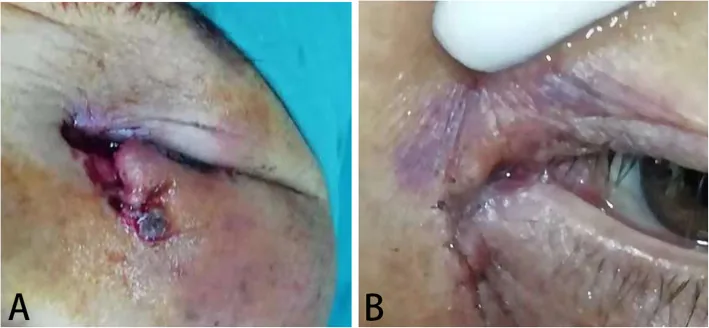
Guo T, et al. Etiology and prognosis of canalicular laceration repair using canalicular anastomosis combined with bicanalicular stent intubation. BMC Ophthalmol. 2020. Figure 2. PMCID: PMC7310031. License: CC BY.
This is a full-thickness eyelid laceration of the left eye with lower canalicular rupture: (a) the preoperative medial canthal laceration and canalicular tear, and (b) the condition after silicone tube placement. It corresponds to the full-thickness eyelid injury with canalicular rupture discussed in section 2, Main symptoms and clinical findings.
Depth assessment is the first important step. Determine whether the injury is limited to the anterior lamella of the eyelid (skin and orbicularis oculi muscle) or is a full-thickness laceration reaching the posterior lamella (tarsus, conjunctiva, levator muscle, and Müller muscle). The treatment plan differs greatly.
The main points to assess are shown below.
Whether the eyelid margin is torn: Check the alignment of the lash line and the grey line. It often reaches the tarsus.
A horizontal laceration with protruding preaponeurotic fat and ptosis: This is an important finding suggesting perforation of the orbital septum and injury to the levator aponeurosis.
Displacement of the punctum: If the punctum is shifted outward, suspect a canalicular laceration.
Displacement of the medial canthus, excessive rounding, or looseness: Suggests avulsion of the medial canthal tendon (medial palpebral ligament).
With blunt trauma, full-thickness lacerations severe enough to completely sever the tarsal plate are less likely, and injuries are more likely to tear the canaliculus at the innermost part of the eyelid.
QWhat associated injuries should be especially considered in eyelid lacerations?
A
Be sure to check for three injuries: eye injury (globe rupture, corneal perforation, or scleral laceration), traumatic ptosis, and canalicular laceration. If there is an eye injury, it must be treated before eyelid repair. If a canalicular laceration is missed, it can cause chronic tearing, so medial eyelid lacerations should be evaluated especially carefully.
Dog bites: Especially common in children. Bites from pit bulls and terriers tend to cause larger tissue loss.
Falls and collisions: Common causes include children hitting the corner of a desk or bicycle handlebar, and falls in older adults.
Punches and traffic accidents: The main causes of blunt trauma in adults. They often cause a canalicular laceration at the innermost part of the eyelid.
Windshields and hooks: May cause sharp cuts or full-thickness lacerations.
Birth trauma: Can occur during manipulation at cesarean delivery.
Sex and age: More common in males and in children to young adults.
Drugs and alcohol: Increased injury risk due to impaired judgment.
Work environment: Inexperienced workers, heavy machinery, fast-moving objects, and work around hooks at eye level.
QHow can eyelid lacerations be prevented?
A
For children, supervision is important when playing with dogs or handling sharp objects. Adults should wear protective eyewear or a helmet during ball sports, cycling, and work. Older adults should work to reduce the risk of falls.
Checking the pre-injury condition: compare with the other eye and photos taken before the injury, and use this for preoperative assessment of traumatic ptosis and scarring.
Mechanism of injury, timing, and whether any self-treatment was done: these affect the decision for surgery and the timing of surgery.
General information: check allergy history, the time of the last oral intake, and tetanus immunization status.
In bite injuries: check for rabies (dog bites or injuries in endemic areas) and the perpetrator’s HIV and hepatitis virus status.
In children or when under the influence of drugs: gather information from parents or witnesses, and keep the possibility of abuse or neglect in mind.
First evaluate whether there is an eyeball injury. If the eyelid is markedly swollen and opening the eye is difficult, use a Demar hook to retract the eyelid and observe the eye with a handheld slit lamp. If there is corneal perforation, laceration, scleral laceration, or globe rupture, treat these before eyelid management.
Then evaluate the location and depth of the laceration, whether there is a foreign body, and whether there is tissue loss. Be sure to check for levator muscle rupture and canalicular laceration. If possible, also check for injury to the levator aponeurosis, Müller muscle, and lower eyelid retractor aponeurosis (LER).
Insert a bougie through the punctum to confirm whether there is a laceration
First-line. Perform before anesthesia
Irrigation test
Inject saline through the punctum and check for leakage
Perform carefully (leakage into surrounding tissue can cause swelling and make intraoperative handling difficult)
Among laceration sites, lower canalicular lacerations are most common, followed by upper canalicular lacerations, then lacerations of both canaliculi. In indirect injuries from blunt trauma, the tear occurs on the nasal side, making it harder to find the cut ends than in direct injuries (sharp cuts).
If history and examination suggest a foreign body or orbital fracture, obtain a CT scan.
CT (brain, orbit, face): Evaluate axial, coronal, and parasagittal views with 1-2 mm slices.
MRI (T1-weighted): Useful for visualizing wooden foreign bodies. However, MRI is contraindicated if a metallic foreign body is present. The first-line initial test is CT. Note that wood, plastic, and some glass may not appear on X-ray or CT.
Differentiate from contusion around the eye, avulsion of the canthal ligament, and eyelid avulsion. Also keep in mind associated injuries such as corneal foreign body, orbital fracture, and traumatic hyphema.
Before starting surgery, rule out globe rupture, retained foreign body, orbital fracture, and intracranial injury. If there is an eye injury, it takes priority over eyelid treatment. In principle, primary repair should be performed within 12-24 hours after injury 1. However, in a retrospective study by Chiang et al., cases repaired more than 24 hours after injury did not show a significant increase in complication rates, and delayed repair may be acceptable depending on the situation 3.
Superficial horizontal laceration (for example, injury from the corner of a desk in a child): after disinfection and pressure hemostasis, tape closure alone may be sufficient.
Simple superficial laceration involving less than 25% of the eyelid and following a skin crease: may be managed with triple antibiotic ointment or skin adhesive.
Anesthesia: Perform infiltration anesthesia with 0.5% or 1.0% lidocaine containing epinephrine.
Irrigation: Remove all foreign bodies such as sand, mud, and glass fragments with saline. Fine foreign bodies should be removed under an operating microscope.
Debridement should be kept to a minimum: Only clearly crushed or contaminated tissue should be excised. Eyelid skin has a rich blood supply and is resistant to infection, so even when crush injury is severe, it usually survives well after suturing. Debridement can create tissue loss, so it is better not to perform it.
Hemostasis: Treat arterial bleeding with bipolar electrocautery.
No eyelid margin laceration: Align the wound edges precisely and suture them together.
Skin closure: Use 7-0 nylon suture.
Thick skin of the eyebrow and nasal root: Add buried sutures with 6-0 nylon.
Multiple flap-like lacerations: Although they may look like tissue loss at first, in most cases they can be closed without a skin defect if the wound edges are carefully matched.
Complex laceration (eyelid margin and tarsal plate laceration)
Temporary eyelid margin suturing: First place a temporary suture with 6-0 nylon, secure it with a mosquito clamp, and tension the tarsal plate.
Tarsal plate suturing: Suture the lacerated tarsal plate with 6-0 nylon.
Posterior lamellar reconstruction: Suture the tarsus, then the Müller muscle, then the levator. If the medial or lateral canthal tendon is torn, suture it back in place.
Final eyelid-margin suturing: After skin closure, remove the temporary sutures and resuture so that the lash line and grey line align precisely.
For a full-thickness laceration involving the tarsus, align the wound edges using the meibomian gland openings as a guide, and pass 6-0 nylon from the eyelid margin into the tarsus. After placing 2 to 3 sutures in the anterior surface of the tarsus with 6-0 absorbable suture, realign the eyelid margin with 8-0 absorbable suture.
In patients for whom follow-up is difficult (children, people with dementia, homeless patients), avoid using non-absorbable sutures.
Even if only one canaliculus is torn, canalicular reconstruction is the rule. Surgery within 48 hours after injury is preferable, and it can be performed relatively easily within 1 week. The longer the delay, the more scarring develops and the harder it becomes to find the cut ends. In an analysis of 137 cases by Murchison et al., the success rate of repair in the operating room was 85.9%, whereas the success rate in a minor procedure room was only 36.8%, showing that repair at a specialized center greatly contributes to prognosis4.
Choice of anesthesia: General anesthesia is preferable when a canalicular laceration is present. This is because tissue swelling from local anesthesia makes it difficult to find the cut ends. If the procedure is done under local anesthesia, combine it with an infratrochlear nerve block.
The surgical steps are as follows.
Insert a bougie through the punctum to estimate the site of the laceration.
Use a fishhook and traction suture (such as 4-0 silk) to expose the wound.
Control bleeding with epinephrine-soaked gauze or bipolar cautery while searching for the cut ends. The cut ends of the canaliculus appear as a shiny ring with a milky white to grayish white color.
After identifying the ends, insert a silicone tube through the punctum and pass it into the nasal cavity.
Suture the cut ends of the lacrimal canaliculus from the posterior wall with 8-0 Vicryl or nylon sutures.
Suture not only the lacrimal canaliculus but also the surrounding tissues, including Horner’s muscle.
Before closing, check whether the medial canthal ligament is torn, and repair it if it is.
Remove skin sutures 5 to 7 days after surgery (about 1 week). Leave periocular and eyelid-margin sutures in place for 5 to 10 days.
The schedule when a lacrimal tube is left in place is shown below.
Timing
Procedure
Postoperative
Start steroid eye drops + antibiotic eye drops
About 2 weeks after surgery
Perform the first irrigation test (doing it too early can cause leakage at the laceration site and delay healing)
1–2 months after surgery
Tube removal (usual timing)
For 2–3 months after removal
Continue irrigation checks every 2 weeks
With the bicanalicular stent method, the anatomical success rate is about 95.9% and the functional success rate about 89.6%5. Even with the self-retaining mini-MONOKA monocanalicular stent, the anatomical success rate is about 85.7% and the functional success rate about 92.9%6, and both show good treatment results. Similar results have also been reported in large Asian studies of monocanalicular stents7.
Scars are most noticeable 2–3 months after surgery, but they become less noticeable over 6 months to 1 year. Complete wound healing and scar maturation take 6–12 months.
If a contaminated wound, a dog bite, or a foreign body is suspected, use systemic antibiotics (such as amoxicillin-clavulanate, doxycycline, trimethoprim-sulfamethoxazole, and cephalexin). Depending on the injury, also consider tetanus and rabies prophylaxis.
QIf a canalicular laceration is present, how soon should surgery be done?
A
Surgery within 48 hours after injury is preferable, and within 1 week the cut ends can still be found and sutured relatively easily. The longer time passes, the more scarring develops in the surrounding tissue, making the ends harder to identify. Because the procedure is recommended under general anesthesia, early referral to a specialist center is important.
QHow long does it take for an eyelid laceration scar to become less noticeable?
A
The scar is usually most noticeable 2 to 3 months after surgery, but it gradually becomes less visible over 6 months to 1 year. Complete wound healing and scar maturation take 6 to 12 months. During this period, excessive pulling and exposure to ultraviolet light should be avoided.
6. Pathophysiology and detailed mechanism of onset
The skin of the eyelid is the thinnest in the body and contains very little subcutaneous fat. This gives it both excellent mobility and vulnerability to external force.
Layers of the upper eyelid
Anterior layer: Skin (thin, with no subcutaneous fat) → orbicularis oculi muscle (innervated by the VII cranial nerve, divided into the pretarsal, preseptal, and orbital parts)
Septum: orbital septum (about 10 mm above the eyelid margin) → fat pad (located between the septum and the levator aponeurosis, an important landmark for laceration repair)
Posterior layer: levator aponeurosis (innervated by the oculomotor nerve, about 5 mm above the tarsal plate) → Müller muscle (sympathetic innervation, about 10 mm above the tarsal plate) → tarsal plate → conjunctiva
Vascular structure: Two arterial arches: the marginal arterial arch (about 2 mm above the eyelid margin) and the peripheral arterial arch (at the peripheral edge of the tarsal plate)
Layers of the lower eyelid
Upper 5 mm (tarsal region): Skin → pretarsal orbicularis oculi muscle → tarsal plate → conjunctiva, a four-layer structure
Lower 5 mm (septal region): Skin → preseptal orbicularis oculi muscle → orbital septum → fat pads (nasal, central, and temporal) → capsulopalpebral fascia → inferior tarsal muscle → conjunctiva, a seven-layer structure
Function of the tarsal plate: Maintains the structural integrity of the eyelid and houses the meibomian glands and their openings, as well as the eyelash follicles
The tear drainage pathway is puncta → canaliculi → common canaliculus → lacrimal sac → nasolacrimal duct.
Dimensions of the canaliculi: diameter 1–2 mm, vertical part (the part that runs along the eyelid from the punctum) about 2.5 mm, horizontal part (the part that runs toward the nose) about 8 mm.
Formation of the common canaliculus: In more than 80% of cases, the superior and inferior canaliculi join to form a common canaliculus.
Clinical significance: The inferior canaliculus is considered the main route for tear drainage, and injury to the inferior canaliculus alone can still cause tearing.
Direct injury (cut by a sharp object): The cut site is clear, and finding the ends is relatively easy.
Indirect injury (blunt trauma): The eyelid is pulled excessively outward and torn on the inner side. Compared with direct injury, the canaliculus ruptures more on the nasal side, and tissue distortion makes it difficult to realign the cut ends.
Doğan E, Bahadır Coşkun Ş, Güner Sönmezoğlu B, Alagöz G. Demographic, Etiological, and Clinical Characteristics of Eyelid Lacerations. Turk J Ophthalmol. 2024;54(1):17-22. PMID: 38385316. PMCID: PMC10895165. ↩↩2
Chiang E, Bee C, Harris GJ, Wells TS. Does delayed repair of eyelid lacerations compromise outcome? Am J Emerg Med. 2017;35(11):1766-1767. PMID: 28473278. ↩
Murchison AP, Bilyk JR. Canalicular laceration repair: an analysis of variables affecting success. Ophthalmic Plast Reconstr Surg. 2014;30(5):410-414. PMID: 24777271. ↩
Guo T, Qin X, Wang H, et al. Eiology and prognosis of canalicular laceration repair using canalicular anastomosis combined with bicanalicular stent intubation. BMC Ophthalmol. 2020;20(1):246. PMID:32571261; PMCID:PMC7310031. doi:10.1186/s12886-020-01506-w. ↩
Alam MS, Mehta NS, Mukherjee B. Anatomical and functional outcomes of canalicular laceration repair with self retaining mini-MONOKA stent. Saudi J Ophthalmol. 2017;31(3):135-139. PMID: 28860909. PMCID: PMC5569334. ↩
Lin CH, Wang CY, Shen YC, Wei LC. Clinical Characteristics, Intraoperative Findings, and Surgical Outcomes of Canalicular Laceration Repair with Monocanalicular Stent in Asia. J Ophthalmol. 2019;2019:5872485. PMID: 31341656. PMCID: PMC6636491. ↩
Copy the article text and paste it into your preferred AI assistant.
Article copied to clipboard
Open an AI assistant below and paste the copied text into the chat box.