Canalicular laceration is an injury caused by acute physical trauma to the canaliculus, part of the lacrimal drainage system. The canaliculus is located on the medial side of the eyelid and does not contain a tarsal plate, making it prone to tearing near the medial canthus.
The most common site of laceration is the inferior canaliculus alone, accounting for approximately 71.9% of cases. The superior canaliculus alone accounts for 15.7%, and simultaneous injury to both the superior and inferior canaliculi or the common canaliculus accounts for 12.4%. The mechanism of injury can be classified as indirect or direct.
A comparison of injury patterns is shown below.
Injury Pattern
Mechanism
Characteristics
Indirect injury
Excessive lateral traction of the eyelid due to blunt trauma
Rupture on the nasal side, difficult to repair
Direct injury
Penetrating trauma from glass, metal, etc., or dog bite
Stump location relatively superficial
ICD-10-CM codes are S01.111A for the right eye and S01.112A for the left eye.
QIs lacrimal canalicular laceration more common in the upper or lower canaliculus?
A
Lower canalicular laceration is most common, accounting for approximately 71.9% of cases. Indirect injury (excessive lateral traction of the eyelid during blunt trauma) causes rupture on the nasal side, making repair difficult and surgical difficulty higher than direct injury.
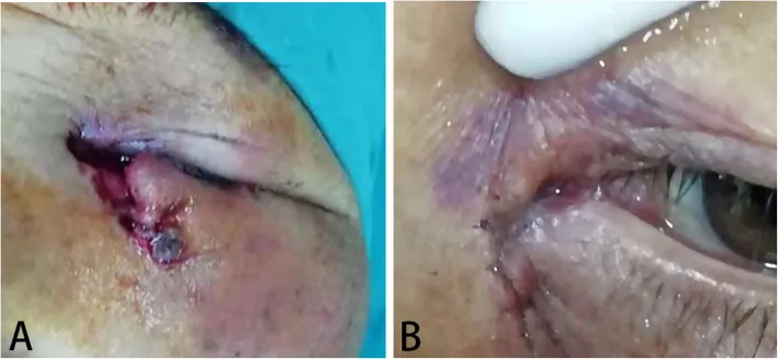
Guo T, et al. Eiology and prognosis of canalicular laceration repair using canalicular anastomosis combined with bicanalicular stent intubation. BMC Ophthalmol. 2020. Figure 2. PMCID: PMC7310031. License: CC BY.
Preoperative image (a) and postoperative image after silicone tube intubation (b) in a female patient with left lower canalicular laceration and full-thickness eyelid laceration, showing tube insertion postoperatively. This corresponds to the lacrimal canalicular laceration discussed in the section “2. Main symptoms and clinical findings.”
Epiphora (tearing): Lacrimal canalicular laceration impairs tear drainage, causing tears to overflow the eyelid margin and run down the cheek (epiphora).
Eyelid laceration medial to the punctum: This is the most important finding and immediately raises suspicion of a canalicular laceration.
Lateral displacement of the punctum: If the medial canthal tendon is also torn, the punctum may be displaced laterally.
Wound gaping: Under a slit lamp, gently pulling the upper eyelid upward and the lower eyelid downward may cause the wound to open, making it easier to identify the cut ends.
Canalicular stump: Appears as a milky white or gray-white, glossy ring.
Bougie insertion findings: Insert a bougie through the punctum and measure the distance to the laceration. A short distance indicates a superficial distal stump, while a long distance suggests the stump is retracted deep near the lacrimal sac.
Irrigation test findings: If leakage is observed, a canalicular laceration can be diagnosed.
Assault/fight injuries: The most common cause of canalicular trauma.
Dog bites: Often occur near the medial canthus, with a canalicular injury rate of 35.6%, significantly higher than the rate from other causes (3.6%). Pit bull terriers are frequently involved.
Blunt shear injuries: Contusions from balls or falls. A major cause alongside assault.
Penetrating trauma: Direct injury from glass, metal, shelf hooks, clothing snaps, etc.
Traffic accidents/falls: The risk of injury from falls increases, especially in the elderly.
Age and sex risk characteristics: most injuries occur in children or young adults. Children under 4 years are more susceptible to facial dog bites, while older adults have a higher risk of falls. Males are more prone to canalicular lacerations than females. Impaired judgment due to alcohol or other substances increases the risk of violence and traffic accidents.
The medial eyelid lacks tarsal plate structure, and anatomical features make it prone to tearing when forces act to detach it from the medial canthal tendon, lacrimal bone, and maxillary bone attachments. Blunt trauma such as punches or ball strikes rarely causes tarsal plate fractures but often results in lacerations with canalicular rupture at the innermost part of the eyelid.
QWhat is the probability of canalicular laceration when bitten on the face by a dog?
A
The rate of canalicular injury from dog bites is 35.6%, approximately 10 times higher than other causes (3.6%). For bites near the medial canthus, canalicular rupture should be strongly suspected, and early evaluation by an ophthalmologist is necessary.
Imaging studies: CT scan if foreign body retention is suspected. MRI is contraindicated if ferromagnetic foreign bodies are present.
Confirmation of complications: Assess for associated globe rupture, corneal perforation, orbital fracture, extraocular muscle injury, and head/face trauma.
Even if only one of the upper or lower canaliculi is torn, lacrimal canalicular reconstruction is the basic approach. The principle of surgery is to restore the separated tissue to its normal structure as much as possible. In lacrimal canalicular rupture, repair is aimed for within the feasible range 1).
Reduction within 48 hours after injury is desirable.
Repair is relatively easy within one week after injury. At the time of injury, it is acceptable to prioritize other treatments with only wound closure and plan for reconstruction at a later date.
Surgery is also indicated for chronic cases, but the more scarring progresses, the more difficult it becomes to find the severed ends.
Successful cases have been reported even at about 72 hours to 5 days after injury, so immediate emergency surgery is not always necessary.
General anesthesia: Preferred for extensive wounds or those involving lacrimal canalicular rupture. Local anesthesia can cause tissue swelling, making it difficult to locate the severed ends.
Local anesthesia: Combined with an infraorbital nerve block. Inject 1–2 mL of 1–2% lidocaine vertically along the orbital wall just above the medial canthal tendon.
Wound exposure: Use skin hooks or traction sutures (e.g., 4-0 silk) to expose the wound and search for the severed ends.
Hemostasis and visualization: Use epinephrine-soaked gauze, bipolar cautery, and suction to achieve hemostasis and clear the field while searching for the severed ends.
Confirmation of the stump: The lacrimal canaliculus stump appears as a gray-white ring. After finding the stump, confirm it is the lacrimal canaliculus by irrigation or bougie insertion.
If the stump is not found: Use low magnification, remove the retractor, and search again by estimating the lacrimal sac side stump based on anatomical location.
② Tube placement
Single canalicular stent: Mini Monoka, etc.
Bicanalicular stent: Use Crawford or Ritleng stents to place from the punctum to the nasal cavity.
③ Canalicular suturing
Stump suturing: Suture with 9-0 to 10-0 nylon or 8-0 absorbable suture, 2 to 3 stitches. Suture from the posterior wall and tie knots outward. Loosen tension before tying.
Peripheral tissue suturing: Suturing surrounding tissues including Horner’s muscle together is important for functional reconstruction.
Medial canthal tendon: Suture if torn (failure leads to lateral punctal displacement and deformity).
Skin suturing: Suture subcutaneous tissue and skin with 7-0 nylon. Do not perform debridement as it causes tissue loss.
④ Postoperative management
Eye drops: Start antibiotic and steroid eye drops from the day after surgery (steroids to prevent foreign body reaction to the tube).
Suture removal: Remove skin sutures after 5 to 7 days (around 1 week).
Irrigation test: Perform for the first time about 2 weeks after surgery (early irrigation may cause leakage at the rupture site and delay healing).
Tube removal: Usually remove after 1 to 2 months, then confirm irrigation every 2 weeks for 2 to 3 months.
Tetanus vaccine: Administer a booster if not vaccinated within the past 10 years.
Rabies management: Consider for animal bites (especially from wild animals).
Prophylactic antibiotics: Recommended for penetrating bites. First-line is amoxicillin/clavulanate (Augmentin). Common causative organisms are Pasteurella canis for dog bites and Pasteurella multocida for cat bites.
QHow soon should surgery for canalicular laceration be performed?
A
Repair within 48 hours of injury is desirable. It can be performed relatively easily within one week. Even in chronic cases, surgery is indicated, but stump identification becomes more difficult as scarring progresses. Immediate emergency surgery is not always necessary; it is acceptable to prioritize the patient’s general condition and other injuries and plan surgery accordingly.
The lacrimal drainage system consists of the punctum, vertical portion (ampulla, about 2 mm), horizontal portion (about 8 mm), common canaliculus (3–5 mm), and lacrimal sac. The punctum is located approximately 6.5 mm from the medial canthus on the upper eyelid and about 6.0 mm on the lower eyelid. The cross-sectional area of the punctum is 0.321 mm² for the lower punctum and 0.264 mm² for the upper punctum, with no statistically significant difference1). The lumen diameter of the canaliculus is about 1–2 mm. The common canaliculus is present in 98% of patients, formed by the union of the upper and lower canaliculi in over 80% of cases.
Contraction of the pretarsal orbicularis muscle: Compresses the ampulla, shortens and compresses the canaliculus. It moves the punctum medially, creating negative pressure to aspirate tears.
Contraction of Horner-Duverney muscle: Applies positive pressure to the lacrimal sac and nasolacrimal duct to push tears into the nasal cavity1).
During eyelid opening
Dilation of the canaliculi and lacrimal sac: Creates negative pressure to aspirate tears1).
Anatomical arrangement of Horner’s muscle: Surrounds the canaliculi in a scissoring pattern, running more densely and parallel around the horizontal canaliculus (findings from electron microscopy and 3D histology)1).
The tear transit time through the upper and lower canaliculi is approximately equal (no statistically significant difference on dacryoscintigraphy), and if one is obstructed, the other can compensate to some extent1).
Clinical significance of unilateral canalicular obstruction
With only unilateral canalicular obstruction, if the eyelid position is normal, tears are effectively drained through the healthy canaliculus. Fewer than 10% of patients with single canalicular obstruction experience epiphora under basal tear conditions1).
It has been reported that 75% of patients with failed lower canalicular repair did not develop epiphora (Ortiz and Kraushar)1). Additionally, Smit and Mourits reported no epiphora in all 16 cases of single canalicular injury that were not repaired1).
Despite the existence of such compensatory mechanisms, repair is pursued whenever possible in canalicular lacerations1). In addition to increased drainage by the healthy canaliculus, an autoregulatory mechanism has been suggested in which punctal occlusion affects the interaction between the ocular surface and lacrimal gland, reducing tear secretion1).
QIf only one canaliculus is severed, will tears always overflow?
A
Epiphora does not necessarily occur. Fewer than 10% of patients with unilateral canalicular obstruction experience epiphora under basal tear conditions, and 75% of failed lower canalicular repairs showed no epiphora1). This is due to compensatory mechanisms by the remaining canaliculus. However, current consensus recommends repair whenever possible.
7. Latest research and future perspectives (research-stage reports)
When a portion of the lacrimal canaliculus remains after tumor resection, a technique to marsupialize the residual canaliculus and use it as a tear drainage pathway is being considered.
In a retrospective study of 22 eyes by Chiu et al., the epiphora rate after marsupialization was 9.1%, showing favorable outcomes compared to the non-reconstruction group (epiphora rate of 12.5% in 97 eyes from the literature)1).
Marsupialization may impair the capillary action and Venturi effect of the lacrimal canaliculus. However, as a simple procedure that can avoid stenosis due to scarring, it is being considered as an alternative in cases where complete repair is difficult1).
Anatomical Elucidation of the Lacrimal Pump Mechanism
Studies using electron microscopy and 3D histology are increasingly clarifying the precise anatomical relationship between the Horner-Duverney muscle and the lacrimal canaliculus. Detailed knowledge of the muscle fiber arrangement around the horizontal canaliculus is expected to contribute to improved accuracy in lacrimal surgery1).
Mohammad Javed Ali, Raman Malhotra, Geoffrey E Rose, Bhupendra C K Patel. Holding back the tears: does marsupialisation of a remnant canaliculus after tumour resection help eliminate epiphora?. BMJ Open Ophth. 2022;7(1):e001090. doi:10.1136/bmjophth-2022-001090.