SLO method (Optomap)

Ultra-widefield fundus camera (Optos and similar)
1. What is a wide-field fundus camera?
Section titled “1. What is a wide-field fundus camera?”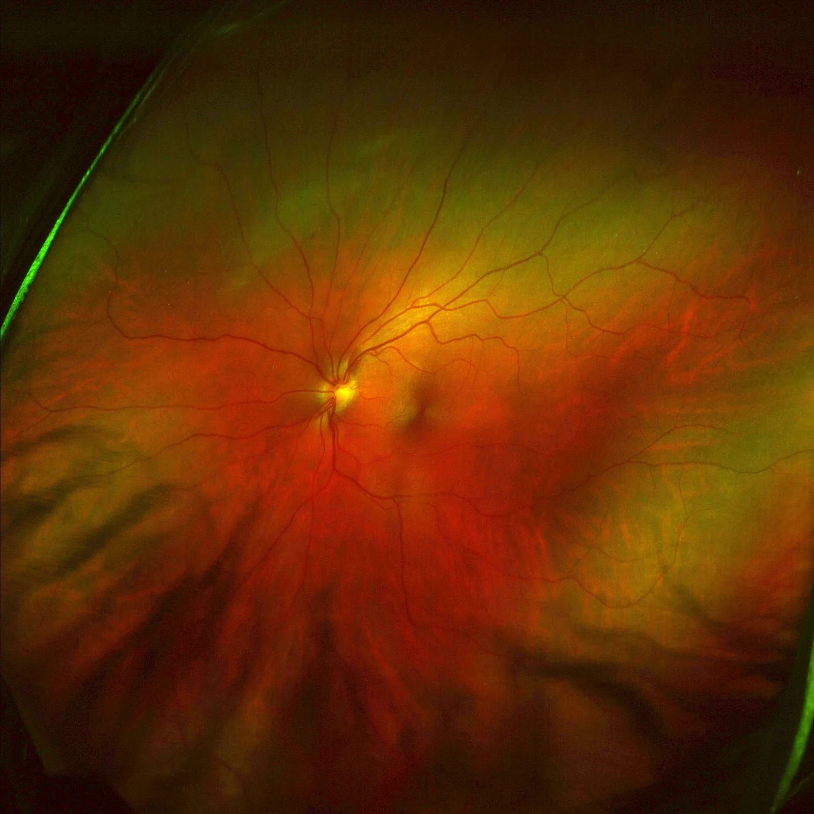
Judgesurreal777 / Overand. Lefteyeoptomap-brightened. Wikimedia Commons. Source: https://commons.wikimedia.org/wiki/File:Lefteyeoptomap-brightened.jpg. License: CC BY-SA 3.0.
This ultra-widefield fundus photograph of the left eye, taken with Optos Optomap, captures more than 200° in a single image, from the optic disc and macula to the peripheral retina beyond the equator. It corresponds to the difference in viewing angle from the standard fundus camera (45°) discussed in the section ‘1. What is a wide-field fundus camera?’.
An ultra-widefield fundus camera (ultra-widefield fundus camera: UWFC) is a device that can image the peripheral retina beyond the equator (more than 200°) in a single shot. Representative models include Optos Optomap and Nidek AFC-330.
A standard fundus camera has a field of view of 45°, and its main area of observation is the posterior pole around the macula and optic disc. With an ultra-widefield fundus camera, the peripheral retina can also be photographed in a single image, allowing broad evaluation of lattice degeneration, retinal breaks, peripheral nonperfusion areas (NPA) in diabetic retinopathy, and peripheral vascular progression in retinopathy of prematurity (ROP).
In settings where non-mydriatic wide-field fundus cameras and OCT are available, fundus findings can be obtained broadly and accurately even without pupil dilation. Depending on the conditions, this may be superior to ophthalmoscopic examination for detecting retinal hemorrhage. However, observation of the most peripheral retinal findings may require a dilated fundus examination.
Ultra-widefield fundus imaging systems can capture the peripheral retina in a single image, but it is important to use them while keeping in mind their limits in distortion and resolution. Reports have shown that detecting peripheral lesions in diabetic retinopathy is associated with an increased risk of retinopathy progression over 4 years1). In addition, widefield fluorescein angiography (widefield FA) has been shown to improve the accuracy of detecting and classifying diabetic retinopathy compared with conventional 7-field ETDRS standard imaging2).
Q
What is the difference between a wide-field fundus camera and a standard fundus camera?
A
Ordinary non-mydriatic fundus cameras have a field of view of about 45°, and mainly photograph the posterior pole centered on the macula and the area around the optic disc. In contrast, ultra-widefield fundus cameras (e.g., Optomap) have a field of view of over 200° and can record areas far out to the periphery beyond the equator of the eyeball in a single shot. They are useful for detecting lesions that ordinary cameras cannot see, such as peripheral retinal tears and degeneration, or peripheral nonperfusion areas in diabetic retinopathy.
2. Classification of imaging methods and representative devices
Section titled “2. Classification of imaging methods and representative devices”Ultra-widefield fundus imaging broadly has two methods, and in clinical practice a three-category classification is used that also includes RetCam, which is specialized for children.
Widefield CCD camera method
Representative device: AFC-330 (Nidek) and others
Light source: White light source
Field of view: about 100–130°
Dilation: usually performed with pupil dilation
Supported modes: color fundus photography; partial FA support
Pediatric wide-angle camera (RetCam)
Representative model: RetCam (Natus Medical)
Field of view: 130° wide-angle lens
Target users: infants and children
Features: A contact wide-angle lens is placed on the cornea for imaging. The drawback is its high cost
Main indications: Screening for ROP (retinopathy of prematurity) and pediatric retinal diseases
| Method | Representative model | Field of view | Need for dilation | Main indications |
|---|---|---|---|---|
| SLO method | Optomap | Over 200° | Not required (recommended) | Diabetic retinopathy, ROP, peripheral retinal degeneration, widefield FA/FAF |
| Widefield CCD method | AFC-330 | 100-130° | Recommended | Peripheral retina observation, color imaging |
| Pediatric contact type | RetCam | 130° | — | ROP, pediatric retinal diseases |
| Standard fundus camera (reference) | Various manufacturers | 45° | Recommended | General posterior pole screening |
3. Indications and clinical significance
Section titled “3. Indications and clinical significance”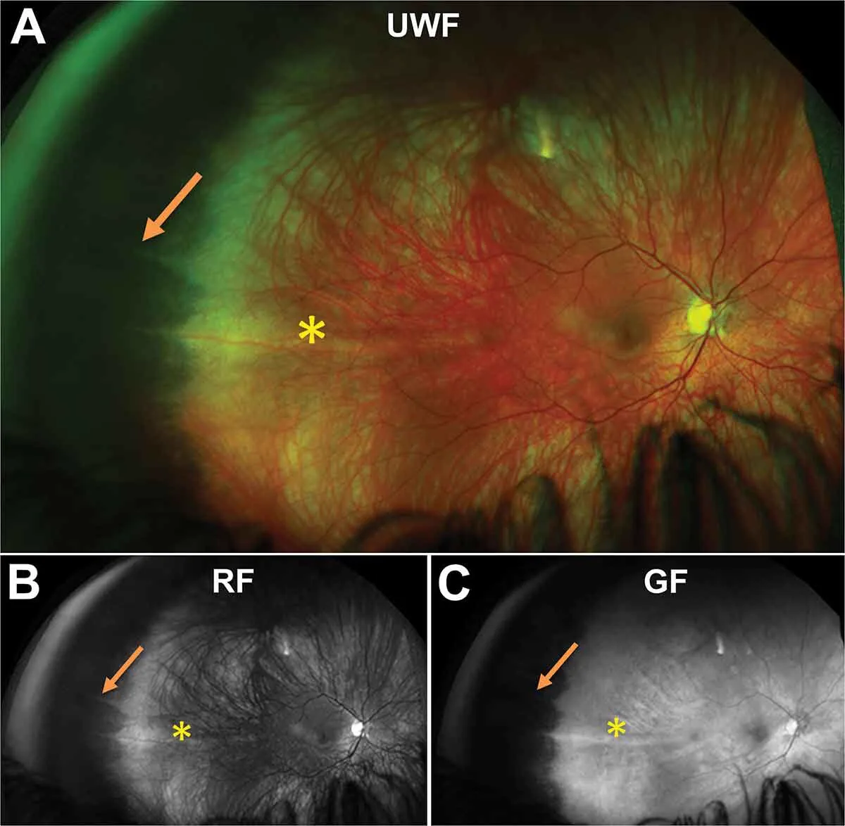
Cheung R, Ly A, Katalinic P, et al. Dentate Processes (Or Ora Tooth). Centre for Eye Health, UNSW Sydney. Wikimedia Commons. Source: https://commons.wikimedia.org/wiki/File:Dentate_Processes_(Or_Ora_Tooth).jpg. License: CC BY 4.0.
.jpg)
Three-panel image captured with an ultra-widefield fundus camera (UWF) showing a dentate process (arrow) near the ora serrata in the temporal peripheral retina (color, red-free, green-free). This corresponds to detection of peripheral retinal lesions discussed in section 3. Indications and clinical significance.
Ultra-widefield fundus cameras are particularly useful for evaluating peripheral lesions that are difficult to capture with standard fundus cameras.
Diabetic retinopathy
Main evaluation items: peripheral NPA (nonperfusion area) and peripheral neovascularization
Clinical significance: the presence of peripheral lesions is associated with the risk of diabetic retinopathy progression over 4 years 1)
Advantages of wide-field FA: Compared with the 7-field ETDRS method, the detection rate for peripheral NPA and neovascularization is improved2)
Application in treatment: Used to determine indications for panretinal photocoagulation (PRP) and for preoperative evaluation6)
Peripheral retinal diseases
Main lesions detected: Peripheral retinal tears, lattice degeneration, snail-track degeneration, and peripheral lesions of retinal vascular occlusion
Clinical significance: Enables early detection of precursor lesions for retinal detachment and helps determine indications for preventive laser photocoagulation
Combined with OCT: Wide-field OCT allows cross-sectional evaluation of the peripheral retina (e.g., Optos Silverstone)3)
Regular follow-up: Also useful for follow-up of postoperative peripheral degeneration
ROP (retinopathy of prematurity)
Device used: RetCam (130° wide-angle contact lens)
Purpose: Screening and periodic evaluation of peripheral vascular development
Implementation: Imaging under general anesthesia or sedation (infants)
Remote screening: The effectiveness of remote screening using RetCam images has been reported5)
Other indications include peripheral choroidal pigment abnormalities, assessment of peripheral RPE degeneration using fundus autofluorescence (FAF), and wide-field evaluation of retinal vein occlusion.
Q
Can the exam be done without dilating the pupils?
A
Optomap can be captured without pupil dilation. It uses natural pupil dilation in a dark room to perform laser scanning, so topical anesthetic drops are not needed either. However, image quality improves when the pupils are dilated, allowing more accurate observation. In particular, when cataracts or a small pupil are present, imaging under dilation may be recommended. RetCam is a contact-type system, so the approach to dilation is different.
4. Examination technique and imaging protocol
Section titled “4. Examination technique and imaging protocol”Optomap imaging procedure
Section titled “Optomap imaging procedure”- Preparation of the environment: Dim the imaging room to encourage natural pupil dilation (when not dilating)
- Positioning: The patient places the chin on the chin rest and looks at the fixation light
- Start scanning: Green and red dual-wavelength lasers scan the retina to obtain a fundus image covering more than 200°
- Peripheral supplemental imaging: Supplemental images of the periphery are acquired by shifting gaze in multiple directions (up, down, temporal, and nasal)
- Add imaging modes: If needed, perform additional imaging in FA, FAF, and ICG modes
When dilation is performed (combined eye drops of 0.5–1% tropicamide and phenylephrine), dilation is complete 15–30 minutes after instillation. Because vision decreases and photophobia occurs for 4–6 hours after dilation, explain this to the patient beforehand. Avoid dilation if angle-closure glaucoma is present or suspected.
RetCam imaging procedure
Section titled “RetCam imaging procedure”- Preparation for sedation or general anesthesia: Because this is for infants and young children, imaging must be done while they remain still
- Probe preparation: Apply methylcellulose gel to the 130° wide-angle lens probe
- Contact imaging: Place the probe against the cornea and carefully capture images all the way to the periphery
- Documentation and assessment: Evaluate the extent of vascular development and whether plus disease is present
Wide-field FA imaging protocol in diabetic retinopathy
Section titled “Wide-field FA imaging protocol in diabetic retinopathy”After intravenous administration of fluorescein (Fluorescite 10% IV 10 mL), obtain wide-field fluorescein angiography in the early phase (30 seconds to 2 minutes) and late phase (10 to 15 minutes). Evaluate the area and distribution of NPA (nonperfusion area) and whether neovascularization is present to determine whether PRP (panretinal photocoagulation) is indicated. The superiority of wide-field FA over the earlier ETDRS 7-field standard photography has been reported2). Confirm any history of contrast allergy and prepare for emergency response.
5. How to read findings and points of caution
Section titled “5. How to read findings and points of caution”When interpreting ultra-widefield fundus images, systematically evaluate the peripheral findings in addition to the posterior pole. Check the peripheral retina in the following four directions in order: superior, inferior, temporal, and nasal.
- Peripheral retinal breaks and lattice degeneration: Check for atrophic breaks and lattice degeneration near the ora serrata
- Peripheral NPA (diabetic retinopathy): Evaluate the extent and distribution of the nonperfusion area that appears white on wide-field FA
- Peripheral neovascularization: In the late phase of FA, confirm fluorescein leakage from peripheral neovascularization on wide-field imaging
- ROP vascular development range: On RetCam images, evaluate the vascular progression line in Zones I to III and plus disease
Q
What should be noted about wide-field fundus photographs?
A
Wide-field fundus photographs, especially images of the periphery, have both distortion and resolution limits. As you move toward the periphery, the image is stretched, so the size and shape of a lesion may look slightly different from reality. In addition, eyelashes or eyelids may appear at the upper and lower edges. If a very peripheral area near the ora serrata looks suspicious, a detailed examination with a dilated fundus examination using a three-mirror lens or indirect ophthalmoscopy is necessary.
6. Technical Principles
Section titled “6. Technical Principles”Principles of the SLO method (Optomap)
Section titled “Principles of the SLO method (Optomap)”Optomap (Optos) uses a scanning laser ophthalmoscope (SLO) system. It generates an ultra-widefield fundus image of more than 200° using the following mechanism.
- Two-wavelength laser: Two lasers are used, green (532 nm) and red (633 nm). Green light is suitable for observing the superficial retina (blood vessels, hemorrhage, nerve fiber layer), while red light is suitable for observing the deeper retina (choroid, RPE)
- Virtual scanning point technology: By scanning the laser from a virtual focal point inside the eye, the peripheral retina can also be scanned while correcting distortion caused by the curvature of the cornea and lens.
- Confocal optics: Detects only reflected light from a specific depth, allowing high-contrast images to be obtained.
- Simultaneous dual-wavelength scanning: Generates pseudo-color images by acquiring 532 nm and 633 nm signals at the same time (the green channel is assigned to the red and green channels, and the red channel is assigned to the blue channel).
This technology makes it possible to obtain ultra-widefield fundus images of more than 200° in a short time (a few seconds) without pupil dilation.
Principles of imaging modes
Section titled “Principles of imaging modes”- Ultra-widefield fundus autofluorescence (FAF): A 488 nm or 532 nm laser excites lipofuscin, allowing broad evaluation of the metabolic state of the peripheral RPE (retinal pigment epithelium).
- Ultra-widefield FA (fluorescein angiography): After intravenous injection of fluorescein, the retinal blood vessels are imaged with a laser. Useful for evaluating peripheral NPA.
- Widefield OCT (Optos Silverstone): In addition to ultra-widefield images obtained by SLO, OCT scans are integrated, making peripheral cross-sectional evaluation possible3)
7. Latest research and future prospects
Section titled “7. Latest research and future prospects”- Peripheral retinal cross-sectional evaluation with ultra-widefield OCT (Optos Silverstone): A device that integrates SLO widefield fundus photography and OCT is making it possible to observe the cross-sectional structure of the peripheral retina, which was previously difficult to evaluate. It has been reported that UWF-OCT with Optos Silverstone can obtain retinal cross-sectional images of more than 100° in a single scan3)
- Automated analysis of ultra-widefield fundus images using AI: A deep learning system has been developed to automatically detect proliferative diabetic retinopathy (untreated cases) from ultra-widefield fundus images. High sensitivity and specificity have been reported, and its future use in screening is expected4)
- Improved accuracy of peripheral NPA assessment with ultra-widefield FA/ICG: Several studies have shown that ultra-widefield fluorescein angiography improves the detection of peripheral NPA and neovascularization in diabetic retinopathy, as well as the accuracy of severity grading, compared with 7-field ETDRS standard photography2). Quantitative assessment of peripheral NPA is expected to help refine the timing of treatment intervention
- Application to tele-screening for ROP: Clinical trials have shown that a tele-screening system using RetCam makes it possible to evaluate retinopathy of prematurity at facilities without an on-site ophthalmologist. The effectiveness of telemedicine systems for assessing acute ROP has been confirmed5)
- Smartphone-linked ultra-widefield fundus photography: Research is also progressing on ultra-widefield fundus photography using smartphone adapters, and its use for ROP and diabetic retinopathy screening in resource-limited settings is being explored
8. References
Section titled “8. References”-
Silva PS, Cavallerano JD, Haddad NM, et al. Peripheral lesions identified on ultrawide field imaging predict increased risk of diabetic retinopathy progression over 4 years. Ophthalmology. 2015;122(5):949-956.
-
Wessel MM, Aaker GD, Parlitsis G, et al. Ultra-wide-field angiography improves the detection and classification of diabetic retinopathy. Retina. 2012;32(4):785-791.
-
Choudhry N, Golber KA, Ferrara D, et al. Ultra-widefield steering-based spectral-domain optical coherence tomography imaging of the retinal periphery. Ophthalmology. 2020;127(9):1272-1274.
-
Nagasawa T, Tabuchi H, Masumoto H, et al. Accuracy of ultrawide-field fundus ophthalmoscopy-assisted deep learning for detecting treatment-naïve proliferative diabetic retinopathy. Int Ophthalmol. 2019;39(10):2153-2159.
-
Quinn GE, Ying GS, Daniel E, et al. Validity of a telemedicine system for the evaluation of acute-phase retinopathy of prematurity. JAMA Ophthalmol. 2014;132(10):1178-1184.
-
日本糖尿病眼学会. 糖尿病網膜症診療ガイドライン(第1版). 日眼会誌. 2020;124(12):955-981.