
Oxymetazoline (Upneeq)
Key Points at a Glance
Section titled “Key Points at a Glance”1. What is Oxymetazoline (Upneeq)?
Section titled “1. What is Oxymetazoline (Upneeq)?”Oxymetazoline hydrochloride 0.1% ophthalmic solution, marketed as Upneeq (RVL Pharmaceuticals), received FDA approval in the United States in 2020. It is the first eye drop to obtain FDA approval as a pharmacological treatment for acquired blepharoptosis.
Target condition is acquired blepharoptosis, particularly involutional (age-related) ptosis. The prevalence of ptosis in adults is reported to be 4.7–13.5%, with incidence increasing with age. Acquired blepharoptosis is a condition in which the upper eyelid gradually droops due to aging, trauma, or muscle dysfunction, affecting both visual field and aesthetics.
Previously, surgical intervention (e.g., levator advancement) was the main treatment. The introduction of Upneeq has added a non-surgical option.
The main classifications of ptosis are as follows.
- Aponeurotic (involutional): Stretching and thinning of the levator aponeurosis due to aging or long-term contact lens wear. This is the most common type.
- Neurogenic: Ptosis caused by oculomotor nerve palsy (e.g., cerebral aneurysm, diabetic ischemia) or Horner syndrome.
- Myogenic: Ptosis due to mitochondrial encephalomyopathies such as chronic progressive external ophthalmoplegia (CPEO).
- Myasthenia gravis (MG) type: Neuromuscular junction disorder. Approximately 70% of initial symptoms of MG are ptosis.
- Pseudoptosis: Apparent drooping due to skin laxity, thyroid eye disease, facial nerve palsy, etc.
2. Main Symptoms and Clinical Findings
Section titled “2. Main Symptoms and Clinical Findings”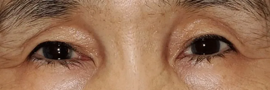
Subjective Symptoms
Section titled “Subjective Symptoms”- Drooping of the upper eyelid: Occurs in one or both eyes.
- Narrowing of the upper visual field: Difficulty seeing upward, interfering with daily activities.
- Compensatory frontalis muscle tension: Attempts to raise the eyebrows lead to increased forehead wrinkles, tension headaches, or deep eye pain.
- Impact on aesthetic appearance: The drooping eyelid appearance may be a concern.
Clinical Findings (Findings Confirmed by Physician Examination)
Section titled “Clinical Findings (Findings Confirmed by Physician Examination)”- Decreased MRD1 (margin reflex distance 1): The distance from the corneal light reflex at the center of the pupil to the upper eyelid margin. Normal is 3.5 mm or more.
- Assessment of levator palpebrae superioris function: Press down on the eyebrow and have the patient look upward, measuring the amount of eyelid elevation. Preserved levator function suggests aponeurotic ptosis.
- Loss or irregularity of the eyelid crease (double eyelid): An indicator of aponeurotic stretching.
- Presence of pupillary abnormalities: Important for excluding oculomotor nerve palsy. If accompanied by mydriasis or anisocoria, suspect neurogenic causes.
- Diurnal variation: If symptoms tend to worsen in the evening, it is important to rule out myasthenia gravis.
3. Causes and Risk Factors
Section titled “3. Causes and Risk Factors”The main causes of acquired ptosis are listed below.
- Involutional (aponeurotic): Stretching and thinning of the levator aponeurosis due to aging. This is the most common cause.
- Long-term contact lens wear: Causes mechanical irritation leading to aponeurotic degeneration.
- Neurogenic: Ptosis due to oculomotor nerve palsy (e.g., cerebral aneurysm, diabetic ischemia). Acute onset may indicate a cerebral aneurysm and requires semi-urgent management.
- Horner syndrome: Damage to the sympathetic efferent pathway results in the triad of blepharoptosis, miosis, and anhidrosis.
- Myogenic (CPEO): Chronic progressive external ophthalmoplegia. Associated with mitochondrial encephalomyopathy.
- Myasthenia gravis: Neuromuscular junction disorder. About 70% of initial symptoms are ptosis, with characteristic diurnal variation (worsening in the evening).
Main risk factors: Aging, long-term contact lens use, trauma, history of surgery (induction of Horner syndrome due to eyelid or neck surgery).
The causes of ptosis are diverse. The most common is age-related aponeurotic ptosis, but it can also be caused by conditions that threaten life or visual function, such as oculomotor nerve palsy, Horner syndrome, myasthenia gravis, and chronic progressive external ophthalmoplegia. Since treatment differs depending on the cause, appropriate differential diagnosis is important.
4. Diagnosis and Examination Methods
Section titled “4. Diagnosis and Examination Methods”Medical History
Section titled “Medical History”- Onset timing (sudden onset may indicate cerebral aneurysm → semi-urgent response)
- Contact lens wear history
- Diurnal variation (to rule out myasthenia gravis)
- History of systemic diseases, trauma, and surgery
Ophthalmic measurements
Section titled “Ophthalmic measurements”- MRD1 measurement: Measure the distance from the corneal light reflex to the upper eyelid margin. Normal is 3.5 mm or more.
- Levator function test: While holding down the eyebrows, have the patient look upward and measure the amount of eyelid elevation.
- LPFT (Leicester Peripheral Field Test): Quantitatively evaluates the upper visual field using points. It was also used for efficacy assessment in clinical trials.
Tests for Differential Diagnosis
Section titled “Tests for Differential Diagnosis”- Ice test: Apply an ice pack to the upper eyelid for 2 minutes; improvement of 2 mm or more is positive (suspected MG). Sensitivity 80–92%, specificity 25–100%.
- Tensilon test: Administer edrophonium chloride (Antilirium®) 10 mg intravenously in 2.5 mg increments and observe improvement in ptosis.
- Upward gaze test: Have the patient gaze upward for 1 minute; if ptosis or diplopia worsens, suspect MG.
- Anti-acetylcholine receptor (AChR) antibody: Positive in approximately 85% of generalized MG cases and less than 50% of ocular MG cases.
- Imaging (CT/MRI/MRA): Useful for excluding oculomotor nerve palsy and orbital lesions, and for detecting cerebral aneurysms.
5. Standard Treatment
Section titled “5. Standard Treatment”Oxymetazoline 0.1% Ophthalmic Solution (Upneeq)
Section titled “Oxymetazoline 0.1% Ophthalmic Solution (Upneeq)”Dosage and Administration: Instill one drop in the affected eye once daily. This is a single-use, preservative-free vial containing oxymetazoline hydrochloride 0.1% per mL. The effect lasts approximately 8 hours after instillation. It provides temporary improvement and is not a curative treatment.
Inactive Ingredients: Calcium chloride, hydrochloric acid, hypromellose, magnesium chloride, potassium chloride, sodium acetate, sodium chloride, sodium citrate, and water.
Contact Lens Use: Remove contact lenses before instillation and wait at least 15 minutes before reinsertion. When used with other eye drops, maintain an interval of at least 15 minutes between administrations.
Clinical Trial Results
Section titled “Clinical Trial Results”The efficacy of Upneeq was validated in two Phase 3 RCTs (total 304 patients, randomized double-blind placebo-controlled, active drug:placebo = 2:1). The primary endpoints were the change in LPFT and the change in MRD1.
The LPFT changes (difference from placebo) in each study are shown below.
| Time Point | Study 1 (Upneeq vs Placebo) | Study 2 (Upneeq vs Placebo) |
|---|---|---|
| 6 hours after day 1 | +5.2 vs +1.5 (difference 3.7) | +6.3 vs +2.1 (difference 4.2) |
| 2 hours after day 14 | +6.4 vs +2.2 (difference 4.2) | +7.7 vs +2.4 (difference 5.3) |
At all evaluation time points, the Upneeq group showed statistically significant improvement compared to the placebo group (p<0.01). MRD1 also showed significant improvement.
Conventional Surgical Treatment
Section titled “Conventional Surgical Treatment”Surgical treatment is positioned as a definitive treatment for aponeurotic ptosis.
- Levator aponeurosis advancement: Standard procedure for aponeurotic ptosis. Repair and refixation of the aponeurosis are performed.
- Ptosis due to oculomotor nerve palsy: Treatment of the underlying disease is the highest priority. If no improvement is seen after six months, surgery should be considered.
- Ptosis associated with Horner syndrome: Müller muscle resection may be an option. This is related to the fact that the Müller muscle is α-adrenergic.
After instillation once daily, the effect lasts for about 8 hours. This is a temporary improvement and not a cure, so continuous instillation is necessary. Safety and efficacy data for long-term use (over 6 weeks) are outside the scope of clinical trials and remain a future challenge.
Contact lenses should be removed before instillation, and you must wait at least 15 minutes before reinserting them. When used with other eye drops, also allow an interval of at least 15 minutes between administrations.
6. Pathophysiology and Detailed Mechanism of Onset
Section titled “6. Pathophysiology and Detailed Mechanism of Onset”Mechanism of action of oxymetazoline
Section titled “Mechanism of action of oxymetazoline”Oxymetazoline is an alpha-adrenergic receptor agonist (alpha agonist). It binds to alpha receptors on the Müller muscle in the upper eyelid, promoting muscle contraction and thereby lifting the eyelid.
Anatomy and physiology of the Müller muscle
Section titled “Anatomy and physiology of the Müller muscle”The Müller muscle is a sympathetically innervated smooth muscle located deep to the levator palpebrae superioris muscle. Normally, it contributes about 2 mm of eyelid elevation. In Horner syndrome, the sympathetic efferent pathway is disrupted, reducing Müller muscle function and causing blepharoptosis.
Pathophysiology of Acquired (Aponeurotic) Ptosis
Section titled “Pathophysiology of Acquired (Aponeurotic) Ptosis”Stretching and thinning (fibrosis) of the levator aponeurosis reduces transmission of lifting force to the tarsal plate. Traction on the skin via aponeurotic perforators also decreases, causing the eyelid crease to disappear or become irregular. As a compensatory response, the frontalis muscle contracts, leading to eyebrow elevation, forehead wrinkles, and tension-type headaches.
Why Oxymetazoline Is Effective for Involutional Ptosis
Section titled “Why Oxymetazoline Is Effective for Involutional Ptosis”In involutional ptosis, the levator aponeurosis is primarily affected, but Müller’s muscle function often remains intact. Enhanced contraction of Müller’s muscle via alpha-receptor stimulation provides compensatory elevation. This is based on the same principle as why Müller’s muscle resection is effective for ptosis associated with Horner syndrome.
7. Latest Research and Future Prospects
Section titled “7. Latest Research and Future Prospects”Currently published data are mainly based on clinical trials at the time of approval (up to 14 days). The following points have been identified as future challenges.
- Application in elderly: No significant differences in safety and efficacy were observed between elderly patients aged 65 years and older and younger adults.
- Long-term safety: Safety and efficacy data for long-term use exceeding 6 weeks are outside the scope of clinical trials, and evidence is insufficient.
- Application in specific populations: Safety data in pregnant women, nursing mothers, and children under 13 years of age have not been established. In animal studies, oxymetazoline was detected in the milk of lactating rats.
8. References
Section titled “8. References”-
Slonim CB, Foster S, Jaros M, et al. Association of Oxymetazoline Hydrochloride, 0.1%, Solution Administration With Visual Field in Acquired Ptosis: A Pooled Analysis of 2 Randomized Clinical Trials. JAMA Ophthalmol. 2020;138(11):1168-1175. doi:10.1001/jamaophthalmol.2020.3812. PMID: 33001144
-
Bacharach J, Wirta DL, Smyth-Medina R, et al. Rapid and Sustained Eyelid Elevation in Acquired Blepharoptosis with Oxymetazoline 0.1%: Randomized Phase 3 Trial Results. Clin Ophthalmol. 2021;15:2743-2751. doi:10.2147/OPTH.S306155. PMID: 34211263
-
Wirta DL, Korenfeld MS, Foster S, et al. Safety of Once-Daily Oxymetazoline HCl Ophthalmic Solution, 0.1% in Patients with Acquired Blepharoptosis: Results from Four Randomized, Double-Masked Clinical Trials. Clin Ophthalmol. 2021;15:4035-4048. doi:10.2147/OPTH.S322326. PMID: 34675472
-
Newland M, Eberly H, Ma C, Lighthall JG. The Use of Oxymetazoline 0.1% Ophthalmic Solution for Acquired Blepharoptosis: A Systematic Review. Laryngoscope. 2025;135(1):8-14. doi:10.1002/lary.31723. PMID: 39172003
-
Taha M, Li Y, Morren J. Oxymetazoline Hydrochloride Eye-Drops as Treatment for Myasthenia Gravis-Related Ptosis: A Description of Two Cases. Cureus. 2023;15(3):e36351. doi:10.7759/cureus.36351. PMID: 37082493
-
Sung J, Song A, Song M, Song J. Oxymetazoline hydrochloride ophthalmic solution, 0.1%, boosts the effects of botulinum toxin on blepharospasm: a case series. J Med Case Rep. 2022;16(1):299. doi:10.1186/s13256-022-03493-6. PMID: 35927744