Bickerstaff Brainstem Encephalitis (BBE) is a rare autoimmune disease that affects the peripheral nervous system and central nervous system (brainstem). It was first described by Bickerstaff & Cloake in 1951, and Bickerstaff alone later reported it as “a grave syndrome with benign prognosis” in 1957.
Typically, 1 to 4 weeks after a preceding infection, the triad of external ophthalmoplegia, ataxia, and impaired consciousness appears acutely or subacutely. It belongs to the same anti-GQ1b antibody syndrome spectrum as Guillain-Barré syndrome (GBS) and Miller Fisher syndrome (MFS), and is positioned as a subtype of immune-mediated polyneuritis.
A nationwide survey in Japan estimated an annual incidence rate of 0.078 per 100,000 population, with approximately 100 new cases per year4). The male-to-female ratio is 1.3 (slightly male predominant), and the median age of onset is 35 years (mean 39 years)4).
QHow rare is Bickerstaff brainstem encephalitis?
A
A nationwide survey in Japan estimated an annual incidence rate of 0.078 per 100,000 population, with approximately 100 new cases per year4). It is even rarer than Miller Fisher syndrome.
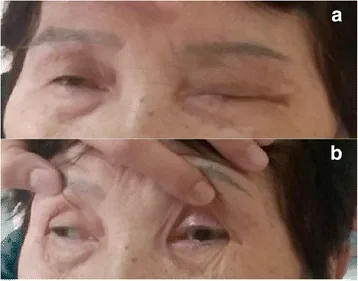
Pei-Ru Chen et al. Posterior reversible encephalopathy as the first manifestation of Bickerstaff’s brainstem encephalitis. BMC Neurology. 2016 Nov 8; 16:215. Figure 1. PMCID: PMC5100286. License: CC BY.
Figure: Clinical findings in Bickerstaff brainstem encephalitis
Pupillary abnormalities: Mydriasis, delayed light reflex, etc., may occur.
Ataxia and Muscle Weakness
Ataxia: One of the three cardinal signs present in all cases.
Limb muscle weakness: Flaccid, symmetric muscle weakness occurs in 60% of cases5).
Hyporeflexia/areflexia: Observed in 58% of cases5).
Hyperreflexia: Conversely, 34% show hyperreflexia, suggesting pyramidal tract involvement4)5).
Babinski sign: Upgoing plantar reflex is positive in 40% of cases4)5).
Consciousness and Brainstem Symptoms
Impaired consciousness: Somnolence in 45%, stupor to coma in 29% of cases4).
Facial nerve palsy: Observed in 45% of cases 6).
Bulbar palsy: Dysarthria and dysphagia occur in 34% 6).
Deep sensory impairment: Observed in 16% of cases 4).
QWhat is the difference between Fisher syndrome and Bickerstaff brainstem encephalitis?
A
Fisher syndrome (MFS) is a disease primarily involving peripheral neuropathy, characterized by the triad of external ophthalmoplegia, ataxia, and areflexia. BBE is distinguished by the addition of consciousness disturbance and pyramidal tract signs (hyperreflexia, positive Babinski sign), indicating more prominent involvement of the central nervous system (brainstem). The two form a continuous spectrum, and overlapping types also exist.
The structure of the pathogen from a preceding infection resembles GQ1b ganglioside, inducing cross-reactive anti-GQ1b antibodies (molecular mimicry). These antibodies are thought to target cranial nerve terminals, dorsal root ganglia, and the blood-brain barrier, causing neurological damage.
Diagnosis is primarily based on clinical findings. The clinical diagnostic criteria by Odaka et al. (2003) are “progressive, relatively symmetric external ophthalmoplegia + ataxia + impaired consciousness or hyperreflexia within 4 weeks”6).
Anti-GQ1b IgG antibody: The most important supportive test. The positivity rate in BBE is 68–80%, and a negative result does not rule out BBE2)5). Sensitivity 60–70%, specificity >90%3). In MFS, it is even higher, with 83–100% positive.
Anti-GM1 antibody: Positive in about 10% of BBE cases2).
Anti-GD1a antibody: Positive in about 13% of BBE cases2).
CSF findings are relatively normal in two-thirds of patients1). Albuminocytologic dissociation (albumin-cytologic dissociation) is observed in about 25% of cases, which is less frequent than in GBS5). CSF protein elevation may progress over time.
Brain MRI is normal in two-thirds of cases. When abnormal, typical findings on T2/FLAIR are hyperintensities in the brainstem (pons and pontomesencephalic junction)2)3)6). FLAIR, T2, and coronal views are useful for detecting brainstem lesions. Contrast enhancement is often absent.
EEG abnormalities are found in 57–70% of cases5)6). Diffuse delta activity is characteristic1)6) and is useful for excluding non-convulsive status epilepticus2).
Findings often overlap with the GBS spectrum. Prolonged or absent F-waves (suggesting early demyelination)6), motor axonal polyneuropathy4), and reduced sensory nerve action potential (SNAP) amplitudes may be observed5). However, some cases show relatively normal results5).
QCan Bickerstaff brainstem encephalitis be diagnosed even if anti-GQ1b antibody is negative?
A
The positivity rate of anti-GQ1b antibody in BBE patients is 68–80%, and negative cases account for 20–30% or more2)5). BBE is a clinical diagnosis; if the classic triad (external ophthalmoplegia, ataxia, and impaired consciousness or hyperreflexia) is present, the diagnosis is not ruled out even if the antibody is negative2).
Currently, there are no RCTs on the treatment of BBE 5)6). Many cases resolve spontaneously, and the prognosis is considered relatively favorable. Treatment is extrapolated from evidence for GBS.
Intravenous immunoglobulin (IVIg): Recommended for progressive GBS or cases with impaired consciousness. Standard regimen is 0.4 g/kg/day for 5 days 2)3). May slightly hasten recovery, but its effect on final outcome is unclear.
Plasma exchange: Reported effective in cases unresponsive to IVIg, especially in children 6).
Steroid pulse therapy: IV methylprednisolone 1 g/day for 5 days may be used in combination 2)3)6). Some reports suggest it may accelerate recovery, but its role is debated, and Cochrane review could not clearly recommend for or against it 5).
Airway management and mechanical ventilation: Required when consciousness deteriorates or respiratory muscle paralysis occurs 2)4).
Physical therapy: Essential for functional recovery during the rehabilitation phase.
Nutritional management and aspiration prevention: In cases with dysphagia, consider tube feeding and saliva management.
QIs there an established treatment for Bickerstaff brainstem encephalitis?
A
Currently, there are no RCTs for the treatment of BBE5)6). IVIg and plasma exchange are used extrapolated from evidence in GBS, but their impact on final outcomes is unclear. Many cases recover spontaneously, and the prognosis is relatively favorable.
GQ1b ganglioside is abundantly expressed in the paranodes and neuromuscular junctions (NMJs) of the oculomotor (III), trochlear (IV), abducens (VI), glossopharyngeal (IX), and vagus (X) nerves. The paranodal and terminal regions of the oculomotor nerves have a higher density of GQ1b than other cranial nerves, which is the main cause of extraocular muscle palsy. GQ1b accounts for only 5–6% of gangliosides in peripheral nerve tissue, but 11–13% in cranial nerves 5).
It is also abundant in large cells (Ia neurons) of the dorsal root ganglia, leading to ataxia and areflexia due to impaired sensory input.
Nerve terminal damage by anti-GQ1b antibodies: Blockade occurs at both pre- and postsynaptic sites in the terminals of cranial nerves III, IV, and VI.
Dorsal root ganglion Ia neuron damage: Loss of sensory input leads to ataxia and areflexia.
Node-paranodopathy: A new concept proposing the Ranvier node as the site of dysfunction. A pattern of damage that is neither “demyelinating” nor “axonal” 5).
Central nervous system damage mechanism (specific to BBE)
Blood-brain barrier (BBB) disruption: BBE serum increases mucocutaneous pemphigoid secretion in brain microvascular endothelial cells (BMECs), disrupting the BBB (in vitro study by Saito et al. 2013) 4)6). MFS serum does not affect the BBB, and the degree of BBB disruption determines the clinical phenotype of Fisher/BBE.
Development of consciousness disturbance: Due to damage to the brainstem reticular activating system 6).
Area postrema pathway: The microcirculation of the area postrema in the brainstem has relatively high permeability to large molecules, serving as a route for antibody entry into the brainstem 6).
In rare autopsy cases, perivascular lymphocytic infiltration, edema, and glial nodules are observed in the brainstem, with diffuse upregulation of HLA-DR-positive macrophages/microglia prominent throughout the brainstem 1). Lesions may also extend to the cerebellar white matter, corpus callosum, spinal gray matter, posterior funiculus, and spinal nerve roots 1).
In the autopsy case by Imam et al. (2022), focal reactive meningothelial proliferation and sparse chronic inflammatory cells were confirmed in the temporal leptomeninges. Immunohistochemistry showed diffuse upregulation of HLA-DR-positive macrophages/microglia throughout the brainstem, also present in the cerebellar white matter, corpus callosum, spinal gray matter, posterior funiculus, and spinal nerve roots 1).
7. Latest Research and Future Perspectives (Investigational Reports)
Pattern changes from anti-GQ1b antibody negativity to positivity for other anti-ganglioside antibodies have been reported at recurrence, and conversely, seroconversion from negative to positive has been reported 12 years after the initial episode 5). Based on observations that anti-GQ1b antibody titers follow the clinical course, the possibility of predicting recurrence through serial anti-ganglioside testing is being investigated 5). The recurrence rate in MFS/BBE is approximately 14%, which is higher than that in GBS (4%) 5).
Integrated Understanding of the BBE-MFS-GBS Spectrum
Multiple studies, including an analysis of 581 cases, have accumulated evidence that BBE, MFS, and GBS form a continuous spectrum 4)5)6). Reports of the Fisher-Bickerstaff overlap syndrome, an overlap between BBE and MFS, are also increasing.
There are reports of rituximab (anti-CD20 monoclonal antibody) being used for refractory cases that do not respond to standard IVIg or plasma exchange, but the evidence remains at the case report level 5).
Based on findings from cases complicated by ulcerative colitis (IBD), it has been suggested that immune dysregulation of the gut-brain axis may be involved in the pathogenesis of BBE 3). The association between non-biologic immunomodulators such as mesalazine and neurological complications is also being investigated 3).
Imam I, Sarrigiannis PG, Shivane AG. Bickerstaff brainstem encephalitis: clinical, neurophysiological, laboratory and postmortem findings of a case presenting as encephalomyelitis. BMJ case reports. 2022;15(2). doi:10.1136/bcr-2021-245588. PMID:35110279; PMCID:PMC8811546.
Warcup A, Movio G, Dhar S, et al. Bickerstaff Brainstem Encephalitis Presenting With Negative Anti-GM1 and Anti-GQ1B Antibodies. Cureus. 2024;16(6):e61653.
Joo H, Lee CS, Joe S, Han J, Kim HK, Cho H. Bickerstaff’s brainstem encephalitis: a rare case of neurologic complication in Ulcerative Colitis. BMC neurology. 2023;23(1):386. doi:10.1186/s12883-023-03430-0. PMID:37884876; PMCID:PMC10601158.
Pantbalekundri N, Acharya S, Shukla S, Kumar S, Malali S. Bickerstaff’s Brainstem Encephalitis and Miller Fisher Syndrome: A Rare Overlap. Cureus. 2024;16(2):e55000. doi:10.7759/cureus.55000. PMID:38550443; PMCID:PMC10973922.
Bhatia SS, Canepa C, Notarianni A. Bickerstaff’s brainstem encephalitis mimicking herpetic encephalomyelitis in a liver transplant patient with anti-GQ1b antibodies. BMJ Case Rep. 2022;15:e251784.
Wong CK, Ng CF, Tan HJ, et al. Bickerstaff brainstem encephalitis with Guillain-Barré syndrome overlap following chlamydia infection. BMJ Case Rep. 2021;14:e242090.
Copy the article text and paste it into your preferred AI assistant.
Article copied to clipboard
Open an AI assistant below and paste the copied text into the chat box.