Miller Fisher Syndrome (MFS) is an autoimmune neuropathy characterized by the triad of acute external ophthalmoplegia, ataxia, and areflexia. It is classified as a subtype of Guillain-Barré syndrome (GBS).
Charles Miller Fisher first described it in detail in 1956. James Collier is said to have already defined the triad in 19321). MFS is part of the anti-GQ1b antibody syndrome spectrum, forming a continuum with GBS, Bickerstaff brainstem encephalitis, and acute ophthalmoplegia (ataxia-free type).
Epidemiology is as follows:
Worldwide prevalence: approximately 1 per 1,000,000 people1)
Incidence: 0.09 per 100,000 people2)
Proportion of GBS: About 5% in Western countries, 17–25% in East Asia1)
Sex ratio: 2:1 male predominance
Mean age of onset: 40 years. Can occur at any age.
Recurrence rate: 11–14%1)
MFS often follows a monophasic and self-limited disease course.
QHow rare is Miller Fisher syndrome?
A
The worldwide prevalence is 1 per million people, and the incidence is 0.09 per 100,000, making it a rare disease1, 2). However, in East Asia, it accounts for 17–25% of GBS cases, which is higher than the approximately 5% in Western countries1).
Juyuan Pan, Ningyu Zheng, Dan Yu et al. Unilateral ophthalmoplegia in anti-GQ1b antibody syndrome: case report and systematic literature review. Frontiers in Immunology. 2025 Oct 10; 16:1669821. Figure 1. PMCID: PMC12549634. License: CC BY.
Illustration showing the three main symptoms of Miller Fisher syndrome: ophthalmoplegia, ataxia, and areflexia
Most cases develop diplopia or unsteadiness (ataxia) about one week after an upper respiratory tract infection. Symptoms progress for 1–2 weeks after onset and then tend to resolve spontaneously.
The representative clinical findings associated with the triad of MFS are shown below.
Ophthalmoplegia
External ophthalmoplegia: Usually bilateral and symmetric. Unilateral involvement is observed in 27–31% of cases4).
Internal ophthalmoplegia (pupillary involvement): Occurs in approximately half of cases. Presents with mydriasis, loss of light reflex, and accommodative dysfunction.
Ptosis: May be observed as a finding outside the triad.
Ataxia
Truncal and limb ataxia: Severity varies. 30% of patients present with ataxia severe enough to prevent independent walking. Unable to perform tandem gait.
Post-onset course: Tends to remit within about 1 month.
Loss of tendon reflexes
Loss of deep tendon reflexes: Considered relatively characteristic of this syndrome. Reflexes may be preserved in 12–31% of cases.
Post-onset course: Recovery tends to be slower than that of ataxia and ophthalmoplegia.
Order of recovery: Ataxia → ophthalmoplegia → areflexia recover in that order1). Ataxia resolves in about 1 month, external ophthalmoplegia in about 3 months, and most cases resolve without sequelae within about 6 months.
MFS/GBS overlap syndrome: 5.6–7.1% of MFS cases have limb weakness2).
Incomplete MFS: Acute ophthalmoplegia without ataxia is recognized as a form of anti-GQ1b antibody syndrome5).
QIn what order do symptoms of Miller Fisher syndrome recover?
A
Recovery proceeds in the order: ataxia (about 1 month) → ophthalmoplegia (about 3 months) → areflexia1). Most patients resolve without sequelae within about 6 months.
Molecular mimicry is the main mechanism of pathogenesis. Lipooligosaccharides of pathogens are structurally similar to human ganglioside GQ1b, inducing cross-reactive anti-GQ1b antibodies.
Preceding infection and pathogens:
Upper respiratory tract infection (76%) is most common. Gastrointestinal infection is also seen in 25%.
Median time from infection to neurological symptom onset is 8 days.
Vaccine-related: Onset has been reported after COVID-19 vaccines (BNT162b2, ChAdOx1, inactivated vaccine), influenza vaccine, and pneumococcal vaccine 5, 6, 7).
The main preceding infections and triggers are summarized below.
Genetic risk factors: An association between HLA-DR2 and recurrent MFS has been suggested4).
QCan Miller Fisher syndrome develop after a cold or vaccination?
A
Preceding upper respiratory infection is observed in 76% of MFS patients. Multiple cases have also been reported after COVID-19 vaccines (mRNA and inactivated vaccines) and influenza vaccination5, 6, 7). However, some reports indicate that the prognosis of post-vaccination MFS is favorable in all cases7).
The diagnosis of MFS is based on clinical diagnosis, confirmed by the triad of ophthalmoplegia, ataxia, and areflexia, and by excluding other diseases.
Serum anti-GQ1b IgG antibody is the most important diagnostic aid.
Test
Sensitivity
Specificity
Serum anti-GQ1b IgG antibody
92%
97%1)
CSF anti-GQ1b antibody
20%
100%1)
Serum anti-GQ1b antibodies are positive in 80–90% of MFS patients.
Serum testing is more sensitive than cerebrospinal fluid testing during the first 3 weeks after onset.
Antibody titers correlate with the severity of ophthalmoplegia.
Cerebrospinal fluid examination: Shows albuminocytologic dissociation (elevated albumin with normal cell count). However, this finding is common to neuroimmune diseases.
Neuroimaging (head MRI): Usually normal. Rarely, nonspecific abnormalities in the cerebellum, middle cerebellar peduncles, or midbrain, or brainstem enhancement may be seen.
MFS is a self-limiting disease, and in cases where respiratory function is preserved, supportive therapy (symptomatic treatment) may be sufficient. Most cases resolve spontaneously and have a good prognosis.
Although no effective treatment has been established, the following immunotherapies are used.
Dosage: 2 g/kg body weight divided over 5 days (400 mg/kg/day × 5 days) 3)
Efficacy: No RCT has been conducted for MFS. Retrospective studies show no major impact on outcomes, but there is a report that it slightly accelerates the onset of recovery from ophthalmoplegia (from 13.5 days to 12.0 days after onset).
Efficacy: Case reports have shown success, but retrospective studies have not demonstrated significant benefit. It is attempted together with immunoadsorption therapy to remove anti-GQ1b antibodies.
Additional use of IVMP: There are case reports of adding methylprednisolone pulse therapy (IVMP) for patients with insufficient recovery from IVIG alone8).
It is a self-limiting disease with a good prognosis, often returning to normal within six months.
Ataxia: resolves in about one month
External ophthalmoplegia: resolves in about three months
Average recovery time: approximately 10 weeks
Residual symptoms: present in up to one-third of cases
Recurrence rate: 3–14%; mortality rate: approximately 4%
QCan Miller Fisher syndrome resolve without treatment?
A
Most cases are self-limiting and resolve spontaneously. IVIG may slightly accelerate recovery of ophthalmoplegia, but large retrospective studies have not shown a significant impact on outcomes. However, if progression to GBS or Bickerstaff brainstem encephalitis occurs, IVIG or plasma exchange is recommended 1).
Lipooligosaccharides of pathogens such as C. jejuni and H. influenzae are structurally similar to ganglioside GQ1b, inducing cross-reactive anti-GQ1b IgG antibodies after infection.
C. jejunicst-II gene: Asn51 polymorphism → anti-GQ1b antibodies → ophthalmoplegia and ataxia / Thr51 polymorphism → anti-GM1 and anti-GD1a → limb weakness (GBS type)
Association with COVID-19 vaccine: SARS-CoV-2 spike protein binds to gangliosides via sialic acid → molecular mimicry 6)
GQ1b is abundantly expressed in the following sites, and each localization explains different clinical symptoms.
Paranodal and terminal regions of oculomotor nerves (III, IV, VI): More abundant than in other cranial nerves. Anti-GQ1b antibodies are the main mechanism involved in extraocular muscle palsy.
Large cells of the dorsal root ganglia (group Ia neurons): Damage to Ia neurons causes sensory ataxia and areflexia due to impaired sensory input.
Ciliary ganglion: Causes internal ophthalmoplegia (pupillary and accommodative disorders).
Presynaptic membrane of the neuromuscular junction (NMJ): Anti-GQ1b binds to the NMJ → complement-dependent massive release of acetylcholine → eventual blockade of neuromuscular transmission. Complement-mediated destruction of axon terminals, surrounding synapses, and Schwann cells also occurs.
GT1a is abundantly expressed in the glossopharyngeal and vagus nerves. In anti-GT1a-positive GBS, cranial nerve palsies (ocular palsy 57%, facial palsy 57%, bulbar palsy 70%) have been reported, with 39% requiring mechanical ventilation 8).
Liang et al. (2022) conducted a scoping review of 10 cases and summarized the clinical features of MFS after COVID-19 vaccination 7).
Mean age 63.5 years, 80% male, median time from vaccination to onset 13 days. CSF albuminocytologic dissociation 88.9%, anti-ganglioside antibody positivity 62.5%. All cases had a good prognosis 7).
In MFS after mRNA vaccine (BNT162b2), the affinity of SARS-CoV-2 spike protein to gangliosides via sialic acid is noted as a pathogenic mechanism 6).
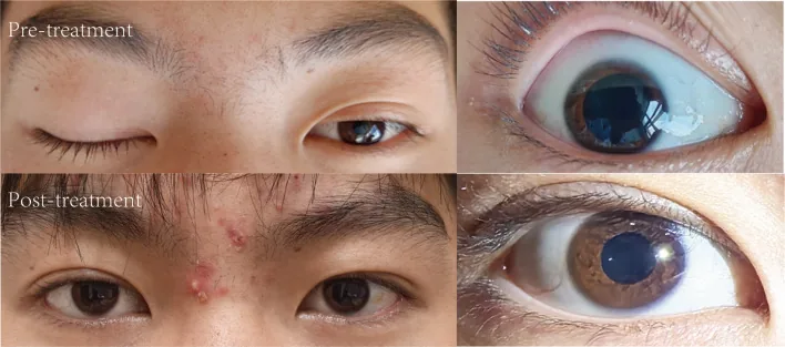
It is known that 27–31% of patients with anti-GQ1b antibody syndrome present with unilateral ophthalmoplegia4).
In a systematic review of 18 cases by Pan et al. (2025), the median age was 31 years, with a male predominance (13/18). Unilateral ophthalmoplegia was observed in 44.4% of children but only 5.7% of adults. Most recovered within 3 months4).
There are case reports of adding IVMP in patients with prolonged recovery after IVIG alone8). A potential effect in promoting recovery has been suggested, but the evidence level remains at the case report stage.
The recurrence rate is reported to be 11–14%, and an association with HLA-DR2 has been suggested1). Recurrences may be milder (e.g., only ophthalmoplegia) than the initial episode, and some cases have been reported to fully recover within one month with conservative treatment alone1).
Ooi ST, Ahmad A, Yaakub A. Recurrent Miller Fisher Syndrome. Cureus. 2022;14(6):e26192. doi:10.7759/cureus.26192. PMID:35891880; PMCID:PMC9306407.
Bahk J, Yang W, Fishman J. Bilateral vocal cord paralysis in Miller Fisher syndrome/Guillain-Barre overlap syndrome and a review of previous case series. BMJ Case Rep. 2021;14:e240386. doi:10.1136/bcr-2020-240386.
Hakobyan N, Yadav R, Pokhrel A, Wasifuddin M, John MJ, Yadav S, et al. Miller-Fisher Syndrome Unveiled in the Presence of Cholangiocarcinoma. Cureus. 2023;15(11):e49016. doi:10.7759/cureus.49016. PMID:38111454; PMCID:PMC10727167.
Pan J, Zheng N, Yu D, Jiang H, Zhou Y. Unilateral ophthalmoplegia in anti-GQ1b antibody syndrome: case report and systematic literature review. Frontiers in immunology. 2025;16:1669821. doi:10.3389/fimmu.2025.1669821. PMID:41142776; PMCID:PMC12549634.
Abičić A, Adamec I, Habek M. Miller Fisher syndrome following Pfizer COVID-19 vaccine. Neurological sciences : official journal of the Italian Neurological Society and of the Italian Society of Clinical Neurophysiology. 2022;43(3):1495-1497. doi:10.1007/s10072-021-05776-0. PMID:34817727; PMCID:PMC8611397.
Yamakawa M, Nakahara K, Nakanishi T, Nomura T, Ueda M. Miller Fisher Syndrome Following Vaccination against SARS-CoV-2. Internal medicine (Tokyo, Japan). 2022;61(7):1067-1069. doi:10.2169/internalmedicine.8851-21. PMID:35370249; PMCID:PMC9038467.
Liang H, Cao Y, Zhong W, Ma Z, Liu J, Chen H. Miller-Fisher syndrome and Guillain-Barre syndrome overlap syndrome following inactivated COVID-19 vaccine: Case report and scope review. Human vaccines & immunotherapeutics. 2022;18(6):2125753. doi:10.1080/21645515.2022.2125753. PMID:36315834; PMCID:PMC9746535.
Mitsuhashi S, Suzuki A, Hayashi K, Sato M, Nakaya Y, Takaku N, et al. Miller-Fisher Syndrome Following Influenza A Infection. Cureus. 2024;16(3):e56064. doi:10.7759/cureus.56064. PMID:38618457; PMCID:PMC11009552.
Ángel-Páez JA, Hurtado-Bugna S, Aragón-Mendoza RL, Altman-Restrepo M, Díaz-Yamal IJ, Centanaro-Meza GA. Miller Fisher syndrome treated with plasmapheresis during pregnancy: Case report and review of the literature. Revista colombiana de obstetricia y ginecologia. 2021;72(2):210-218. doi:10.18597/rcog.3611. PMID:34506707; PMCID:PMC8425356.
Copy the article text and paste it into your preferred AI assistant.
Article copied to clipboard
Open an AI assistant below and paste the copied text into the chat box.