Low-power lens
+14D to +18D: High magnification, narrow field. Suitable for detailed observation of the posterior pole.

Ophthalmoscopy is a routine examination method for observing the fundus, and is broadly divided into direct and indirect ophthalmoscopy.
Direct ophthalmoscopy provides an upright image at about 15× magnification. In contrast, indirect ophthalmoscopy provides an inverted image at 2–5× lower magnification, but offers a wider field of view and is excellent for observing the peripheral retina.
Binocular indirect ophthalmoscopy (BIO) achieves three-dimensional (stereoscopic) observation of the fundus by projecting the optical axis and the left and right visual axes through the pupil. Unlike monocular indirect ophthalmoscopy, it allows stereopsis because both eyes are used.
The main features of BIO are as follows.
It is useful for observing general fundus diseases, and is particularly effective for three-dimensional evaluation of retinal detachment, assessment of macular edema, and retinal neovascularization. The American Academy of Ophthalmology (AAO) Preferred Practice Pattern 2025 also recommends binocular indirect ophthalmoscopy with scleral depression under dilated pupils for evaluation of acute posterior vitreous detachment, retinal tears, and lattice degeneration[2].
The examination itself is not painful. You may feel a slight irritation when the dilating eye drops are applied. If scleral depression is performed, there may be a mild pressure sensation around the eye, but it is not severe pain.
BIO consists of three components: a headband, a binocular lens with mirrors, and a light source.
The optical principle of indirect ophthalmoscopy is as follows. Light from the source enters the pupil, and the reflected light from the fundus is focused in front of the eye by a convex lens (condensing lens). The examiner observes this image with both eyes.
Magnification is calculated as “refractive power of the eye ÷ refractive power of the condensing lens.” For example, when using a +20D lens, it is 60 ÷ 20 = 3×. The higher the diopter, the lower the magnification and the wider the field of view.
The commonly used condensing lens range is +14D to +30D.
Low-power lens
+14D to +18D: High magnification, narrow field. Suitable for detailed observation of the posterior pole.
Standard lens
+20D: 3x magnification. The most widely used standard lens for adult BIO.
High-power lens
+25D to +30D: Low magnification, wide field. Suitable for use in children, premature infants, and cases with small pupils.
The position of the condensing lens directly affects observation quality. If too close, illumination of the peripheral retina is insufficient; if too far, reflected light from the periphery does not reach the examiner. A holding distance of about 5 cm from the patient’s eye is a guideline.
Stereopsis is achieved by aligning the optical axis and both visual axes into the pupil. Narrowing the interpupillary distance facilitates insertion into the pupil, while widening enhances stereopsis. When evaluating the border of a shallow retinal detachment or macular edema, a slightly enhanced stereoscopic setting is useful.
The following filters are used depending on the purpose.
White light
No filter: Used to observe the overall fundus with natural colors.
Yellow
Yellow filter: Reduces light intensity. Used for patients complaining of photophobia.
Red-free
Red-free filter: Useful for improving visualization of blood vessels, hemorrhages, and nerve fiber layer defects.
Blue
Blue filter: Used for observing lesions of the internal limiting membrane and preretinal layers, and for fluorescein angiography.
Adequate mydriasis is necessary to observe the peripheral fundus. Since the bright light of BIO tends to constrict the pupil, maximal dilation is important.
The mydriatic agents used are as follows:
Combining both agents enhances the mydriatic effect. The effect lasts for several hours after dilation, during which glare and blurred near vision occur.
The standard procedure is as follows.
The reasons why the supine position is recommended are as follows.
The curvature of the anterior part of the eye prevents observation of the far periphery. Scleral depression is a technique that indents the sclera from the outside to bring the peripheral retina into the viewing field.
Instruments: Various depressors such as Schepens, O’Connor, Schocket double-ended, Josephberg-Besser, and Flynn are used.
Application by site:
Situations where scleral depression is especially recommended:
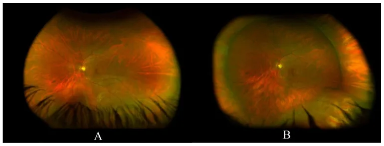
BIO with scleral indentation is considered the gold standard for detecting peripheral retinal tears. Reports indicate that approximately 11% of acute horseshoe tears are missed with non-contact slit-lamp examination, making BIO with scleral indentation essential for evaluating the far periphery [3,5]. In recent years, comparisons with ultra-widefield fundus imaging (UWF) have been conducted, but some reports show that about half of horseshoe tears are missed by UWF, suggesting that UWF alone cannot fully replace scleral indentation [4].
During scleral indentation, intraocular pressure transiently increases significantly, with reports indicating an average of about 65 mmHg (maximum 88 mmHg) even in routine outpatient examinations. Since this may affect ocular perfusion, care should be taken regarding the duration and force of indentation in patients with ocular hypertension or glaucoma [6].
Dilation is necessary to adequately observe the peripheral fundus. After dilation, glare and blurred near vision persist for several hours, so patients should be advised to avoid driving on the day of the examination. In emergencies or when combined with slit-lamp examination using a pre-corneal lens, it may be performed without dilation depending on the purpose.
It is particularly recommended for patients with photopsia or floaters, or those at risk of retinal tears or detachment. Indentation is essential for evaluating the far periphery (near the ora serrata), and tears at the edge of lattice degeneration may only become apparent with indentation.
Advantages:
Disadvantages:
The main differences between monocular and binocular indirect ophthalmoscopy are shown below.
| Item | Monocular indirect ophthalmoscopy | Binocular indirect ophthalmoscopy |
|---|---|---|
| Ease of use | Simple | Complex |
| Stereopsis | Not possible | Possible |
| Scleral depression | Unsuitable | Suitable |
The main differences between direct and indirect ophthalmoscopy are shown below.
| Item | Direct ophthalmoscope | Indirect ophthalmoscope |
|---|---|---|
| Magnification | Approximately 15x | 2–5x |
| Field of view | Narrow (8–10°) | Wide |
| Peripheral observation | Difficult | Easy |
BIO and slit-lamp microscopy (slit-lamp examination) have different roles and are used complementarily.
In clinical practice, the standard procedure is to first perform fundus charting with BIO, followed by detailed examination of the retina and vitreous using a slit lamp with a Goldmann three-mirror lens or similar.
Fundus sketching (fundus charting) using BIO is an important clinical skill.
Fundus charts are essential for managing fundus diseases, especially retinal detachment. It is said that “retinal detachment surgery without a sketch is as reckless as sailing without a chart.” In scleral buckling surgery, the quality of the sketch directly affects surgical outcomes, the surgeon’s skill improvement, and the team’s shared understanding of the pathology.
Chart paper: Retinal detachment charts designed by Schepens and Tolentino are widely used. Typically, three concentric circles (equator, ora serrata, posterior edge of ciliary processes) are printed.
Color code (AAO recommended): Eight colors are used to record retinal findings.
Binocular indirect ophthalmoscopy excels in wide-field stereoscopic observation and is suitable for grasping the positional information of the entire fundus. Slit-lamp biomicroscopy with a pre-corneal lens is superior for detailed observation of vitreoretinal adhesions, etc. The two are complementary; the standard procedure is to create a fundus chart using indirect ophthalmoscopy and then perform detailed examination with the slit lamp.
The history of fundus observation dates back to the 19th century.
Schepens not only developed the binocular indirect ophthalmoscope but also greatly contributed to the popularization of fundus charts and the systematization of retinal detachment surgery. He is called the “father of retinal detachment”[1].