Conservative Treatment
Lacrimal sac massage: Crigler method. Success rate over 85%.
Antibiotic eye drops: Used only when there is excessive discharge.
Indications: First-line treatment until 6 months of age.
Congenital nasolacrimal duct obstruction (CNLDO) is a congenital blockage of the lacrimal drainage system primarily caused by membranous obstruction at the valve of Hasner at the distal end of the nasolacrimal duct. It occurs in 6–20% of newborns and is the most common lacrimal disorder in children 1). The lacrimal duct develops from the lacrimal cord, which forms when ectoderm is buried in the nasolacrimal groove between the lateral nasal process and the maxillary process during embryonic development. The membranous tissue at the lower end of the nasolacrimal duct begins to disappear around the 32nd week of gestation, but remains in about 20% of cases even at the 38th week, just before birth.
Approximately 80% of cases are unilateral, and no sex difference or clear genetic predisposition has been identified 1). The lower opening of the nasolacrimal duct opens at 8 months of gestation, and this condition corresponds to a state where the opening remains incomplete at birth.
The spontaneous resolution rate is high: about 70% resolve by 3 months of age, 80% by 6 months, and 80–100% by 12 months. The spontaneous resolution rate within the first year of life is reported to be 89–96% 1).
89–96% of cases resolve spontaneously within the first year of life 1). Lacrimal sac massage can promote resolution. If symptoms persist beyond 1 year of age, interventions such as probing are considered.
The main symptoms are tearing and eye discharge that appear from early infancy.
The most common cause of CNLDO is membranous obstruction due to delayed regression of the valve of Hasner. Other causes include:
Risk factors for CNLDO are as follows:
Among severe CNLDO cases, complete obstruction 35%, punctal agenesis 15%, congenital fistula 10%, and craniofacial bone defects 5% have been reported 1).
Dacryocystocele is a condition where both the upper and lower parts of the nasolacrimal duct are obstructed simultaneously, causing dilation of the lacrimal sac. It is associated with intranasal cysts in 11–24% of cases and dacryocystitis in 20–74%.
In Down syndrome, CNLDO occurs in up to 30% of cases, which is more frequent than in the general newborn population (6–20%) 1). This is thought to be related to associated craniofacial structural abnormalities.
The diagnosis of CNLDO is based on clinical findings and medical history. The main examination methods are shown below.
| Examination method | Characteristics |
|---|---|
| FDDT | Sensitivity 90%, specificity 100% |
| Lacrimal irrigation test | Confirmation of obstruction |
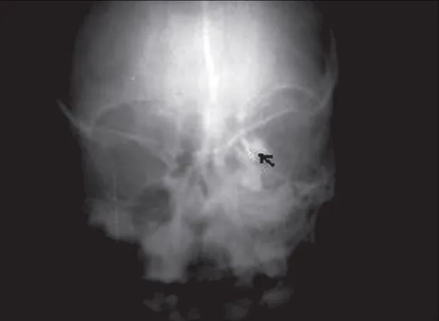
| CT | Evaluation of bony obstruction |
The following diseases must be differentiated.
Conservative treatment is the first choice until 6 months of age.
This procedure involves pressing the area between the inner canthus and nasal root, as well as the nasal ala, with a finger to increase pressure within the nasolacrimal duct and rupture the membrane of the valve of Hasner. The success rate is reported to be over 85% 1).
If there is a large amount of eye discharge, use 0.3% tosufloxacin ophthalmic solution 4 times a day. However, avoid long-term use of antibiotics; use only during periods of heavy discharge.
In Japan, probing is recommended from 6 months of age onward. Before 6 months, it is not recommended due to the risk of sepsis, but early intervention may be considered exceptionally in cases complicated by blepharitis, acute dacryocystitis, or dacryocystocele.
The procedure involves inserting a 0.5-0.6 bougie from the punctum, pulling the eyelid laterally to straighten the canaliculus, advancing about 10 mm to reach the lacrimal sac, and then perforating the membranous obstruction. Insertion from the upper canaliculus is advantageous, as there is less resistance before the sac and a lower risk of damaging the canaliculus. Bending the tip of the bougie approximately 15 degrees at the 10 mm mark helps accommodate variations at the junction of the lacrimal sac and nasolacrimal duct. If restraint is difficult after 1 to 1.5 years of age, the procedure is performed under general anesthesia.
The success rates of probing by age are shown below.
| Age | Success Rate |
|---|---|
| 0–6 months | 90.67% |
| 6–12 months | 85.18% |
| 12–24 months | 82.34% |
| 24–48 months | 85.33% |
| Over 48 months | 63.47% |
(Meta-analysis of 17 studies, 7110 eyes) 3)
The best outcomes are seen in children under 12 months, especially when performed under general anesthesia (0–6 months: 95.42% under general anesthesia, 88.82% under local anesthesia) 3). No significant difference in resolution rate was found between early and late probing (RR 1.00 [95% CI 0.76–1.33]; p=0.99) 2).
The success rate of reprobing decreases to 40–60%. Since iatrogenic canalicular obstruction is found in 44% of cases where initial probing failed, the use of a dacryoendoscope is recommended. Using a dacryoendoscope allows safe opening of the obstruction while visualizing the lacrimal duct lumen, and enables accurate tube insertion under direct vision. If iatrogenic obstruction occurs in the canaliculus, subsequent repair becomes extremely difficult; therefore, if resistance is felt at a site other than the obstruction during probing, the procedure should be stopped and referral to a specialized lacrimal facility should be considered.
Rare complications of probing include bacteremia, meningitis, hip arthritis, and endocarditis. After the procedure, continue antibiotic eye drops and massage.
This is considered when probing is unsuccessful. The success rate of initial stent placement is 90–96%1), and approximately 84% after failed probing. A placement duration of 2 months or longer is recommended.
In complex CNLDO, bicanalicular silicone stents have been shown to be more effective than late probing (RR 0.56 [95% CI 0.34–0.92]; p=0.02)2).
Success rates are reported as 53–95%, but no clear superiority over probing has been demonstrated.
When the above treatments are ineffective or in cases of bony obstruction, dacryocystorhinostomy (DCR) is indicated. Since DCR in children involves invasion of the periosteum, it is generally recommended after around age 15 when facial bone growth is complete, but in severe cases early surgery may be considered. The success rate of external DCR is 96%, and endoscopic DCR is 82–94% 1). Intranasal DCR has the advantages of leaving no facial scar and requiring less bone resection, but requires skilled technique. Postoperatively, a stent is placed for 8–12 weeks. It is the only treatment for bony obstruction.
Conservative Treatment
Lacrimal sac massage: Crigler method. Success rate over 85%.
Antibiotic eye drops: Used only when there is excessive discharge.
Indications: First-line treatment until 6 months of age.
Probing
Technique: Perforate the membrane with a 0.5-0.6 bougie.
Timing: Recommended after 6 months of age in Japan.
Success rate: 63–91% depending on age.
Stenting/DCR
Stent placement: When probing is unsuccessful. Success rate 84–96%.
DCR: For bony obstruction or refractory cases. External approach 96%.
In Japan, probing is recommended after 6 months of age. Up to 16 months of age, there is no significant difference in success rates between early and late probing 2). Since spontaneous resolution is likely up to 12 months of age, conservative treatment centered on massage is generally performed, and probing is considered if there is no improvement.
The success rate of repeat probing decreases to 40–60%. If repeat probing is unsuccessful, stent placement (success rate approximately 84%) is considered, and if that is also ineffective, dacryocystorhinostomy (DCR) is considered.
Lacrimal duct development begins at 3–5 weeks of gestation. The lacrimal groove forms from the surface ectoderm, and canalization progresses around 3 months of gestation 1). The most distal part of the nasolacrimal duct (Hasner’s valve) opens last, usually completing by 8 months of gestation. If this opening is incomplete at birth, CNLDO occurs.
The anatomy of the lacrimal drainage system is as follows.
There are four physiological narrowings of the lacrimal drainage system: the common canaliculus, the lacrimal sac-nasolacrimal duct junction, the nasolacrimal duct, and the nasolacrimal duct opening.
The sites and types of obstruction are as follows:
Sultanbayeva et al. (2025) conducted a meta-analysis of 17 studies involving 7,110 eyes to examine probing success rates by age group 3). For 0–6 months, the overall rate was 90.67% (95.42% under general anesthesia, 88.82% under local anesthesia); for 6–12 months, 85.18% (89.60% under general anesthesia, 82.33% under local anesthesia); for 12–24 months, 82.34% (84.75% under general anesthesia, 75.37% under local anesthesia). Beyond 48 months, the rate dropped to 63.47%. The certainty of evidence was rated low across all subgroups.
A meta-analysis by Farat et al. (2021) of 4 RCTs (423 patients) found no significant difference in resolution rates between early and late probing (RR 1.00; p=0.99; low certainty evidence)2). However, the PEDIG study showed that early probing was more cost-effective ($562 vs $701).
Compared to conventional blind probing, endoscopy-assisted probing is being considered1). The use of dacryoendoscopy allows direct visualization of the obstruction site, potentially reducing the risk of iatrogenic injury and improving success rates.
To improve the success rate of endoscopic DCR, application of mitomycin C to the anastomosis site is being investigated1). It is expected to prevent granulation and scar formation, but long-term safety requires further data accumulation.