Duane Retraction Syndrome (DRS) is a congenital, non-progressive strabismus syndrome first described in detail by Alexander Duane in 1905. It is also called Stilling-Türk-Duane syndrome. It is classified under ICD-10 code H50.81.
It occurs in approximately 1 in 1,000 people in the general population2) and accounts for up to 4% of all strabismus cases. It is the most common congenital cranial dysinnervation disorder (CCDD)2). Unilateral involvement occurs in 82% of cases, more commonly in the left eye (59%), and it is slightly more frequent in females (58%). Bilateral involvement is seen in 15–20% of cases.
Features: Abduction limitation > adduction limitation. May present with esotropia in primary position.
Head posture: Face turn toward the affected side is most common. 72% have a face turn (68% toward the affected side)1)
Epidemiology: Anvari (125 cases) reported Type I in 87.0%1)
Types II and III
Type II (5–10%): Adduction limitation > abduction limitation. Presents with exotropia in primary position and a face turn opposite to that in Type I.
Type III (10–20%): Limited adduction and abduction. 93% have face turn (73% opposite to the affected side) 1)
In a large study (441 patients), face turn was observed in 54.6%, and the frequency of face turn was significantly higher in unilateral cases 1). Horizontal deviation was 76.0%, esotropia 58.4%, and exotropia 17.6% 1).
QHow common is Duane retraction syndrome?
A
It occurs in about 1 in 1,000 people in the general population 2) and accounts for up to 4% of all strabismus. It is the most common among congenital cranial dysinnervation disorders (CCDD). 82% are unilateral, with a slight predominance in the left eye and in girls.
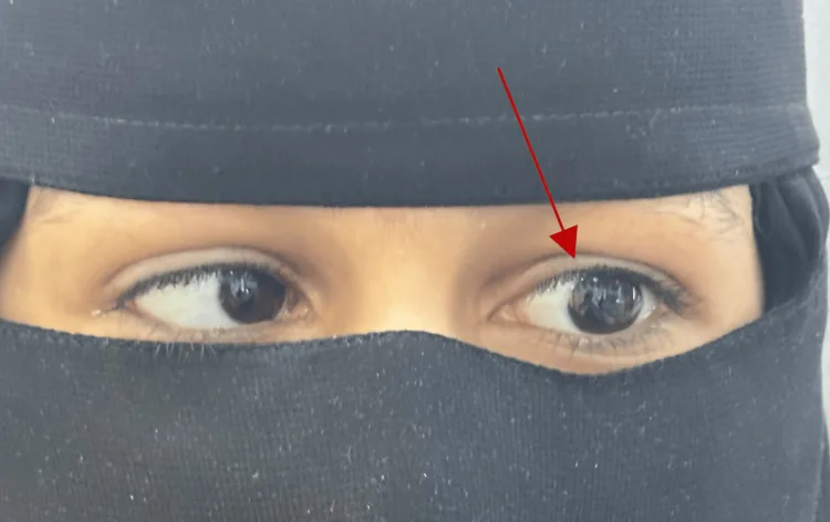
Abu Melha A, Abbas AI, Alghamdi WS, et al. Duane Retraction Syndrome: A Report of Two Cases and Review of Literature. Cureus. 2024 Nov 25;16(11):e74460. Figure 4. PMCID: PMC11680492. License: CC BY.
The left eye does not move sufficiently on lateral gaze, and the abduction limitation is immediately apparent. This clinical photograph shows a typical ocular motility disorder in Duane retraction syndrome.
Abduction limitation: Complete or partial limitation of abduction (degree varies by type).
Globe retraction + palpebral fissure narrowing: On adduction, the globe retracts and the palpebral fissure narrows (induced ptosis). This is a characteristic finding of DRS and is easier to observe from the side.
Widening of the palpebral fissure: The palpebral fissure widens on abduction.
Up-shoot/down-shoot (leash phenomenon): The eye deviates upward or downward on adduction. This is not present at birth but develops secondarily due to progressive contracture of the lateral rectus muscle.
Compensatory head turn is an adaptive movement to maintain binocular vision. If mild (less than 15°), observation without treatment is the principle. Surgery is considered when the turn is 15° or more, or when it causes problems in daily life or cosmesis. At school, environmental adjustments such as seating the child in the direction of the face turn are effective.
Abducens nerve (CN6) deficiency: Damage during the 4th to 8th week of embryonic development leads to absence or hypoplasia of abducens nerve motor neurons.
Abnormal innervation by oculomotor nerve (CN3): The adduction branch of CN3 abnormally innervates the lateral rectus muscle, causing simultaneous contraction of the medial and lateral rectus muscles on adduction, resulting in globe retraction.
Changes in the lateral rectus muscle: Histologically, part of the lateral rectus muscle (the portion not receiving normal innervation) becomes fibrotic.
Associated syndromes: Associated with Duane radial ray syndrome (SALL4 mutation), Goldenhar syndrome, HOXA1 syndrome, etc. 2). About 30% of cases have other congenital anomalies (sensorineural hearing loss, crocodile tears, etc.).
Genetics: 10% of isolated cases are hereditary. Type I is autosomal dominant (8q13), Type II is autosomal dominant (DURS2: CHN1 mutation, 2q31-q32.1). However, 90% are sporadic.
Slit lamp and eye movement examination: Globe retraction and narrowing of the palpebral fissure on adduction are characteristic of DRS. Observing from the side makes it easier to confirm globe retraction.
Forced duction test: Positive (central but with contractile changes in extraocular muscles). Useful for differentiating mechanical restriction from denervation.
MRI: Can depict abducens nerve absence or hypoplasia. Not recommended for routine diagnosis but useful for preoperative evaluation.
Genetic testing (CHN1): Recommended only for familial cases.
Botulinum toxin injection: In a study of 25 patients by Anand, abnormal head posture (AHP) improved from 11.58°±7.43° to 7.86°±6.25° 1). In 16 patients by Ameri, facial turn of 18.27°±7.29° improved to 0.094° after one week, but increased again to 7° after 6 months 1).
Surgical indications: Abnormal head posture of 15° or more, significant deviation in primary position, severe induced ptosis (palpebral fissure height reduced by 50% or more).
Esotropic DRS
Medial rectus recession (4-5 mm) is the basic procedure. Recession beyond 5 mm carries a risk of adduction limitation and consecutive exotropia.
Nishida procedure + medial rectus recession: Esotropia of 40 PD improved to 6 PD, and abduction recovered to 45° 1)
Severe/Refractory Cases
Vertical rectus transposition (VRT): Transposition of the superior and inferior rectus muscles to the lateral rectus insertion. In the augmented suture group, AHP improved from 22.7° to 3.6°; in the non-augmented group, from 18.7° to 7° 1)
Lateral rectus Y-splitting: A procedure for upshoot/downshoot. Combination with rectus recession is also effective 1)
QHow effective is botulinum toxin injection?
A
Botulinum toxin temporarily improves abnormal head posture and esotropia, but the effect diminishes after 6 months 1). It is considered as an alternative or adjunct to surgery for early intervention in infancy or in cases with high surgical risk. Repeated injections may be necessary.
QDoes surgery cure Duane retraction syndrome completely?
A
The underlying neural disorder—congenital absence of the abducens nerve and aberrant innervation by the oculomotor nerve—is not improved by surgery. The goals of surgery are to correct deviation in primary position, reduce compensatory head posture, and improve induced ptosis; normalization of eye movement range is not expected. Many patients can maintain good binocular vision postoperatively through compensatory mechanisms.
The current mainstream theory is the neurogenic theory.
Timing of occurrence: Loss of abducens motor neurons occurs during embryonic weeks 4–8. Postmortem studies at Johns Hopkins University in the 1980s established the neurogenic mechanism.
Mechanism of aberrant innervation: Due to the absence of the abducens nerve, the adduction branch of the oculomotor nerve (CN3) aberrantly innervates the lateral rectus. Simultaneous contraction of the medial and lateral rectus occurs during adduction, leading to globe retraction.
EMG findings: Absence of electrical activity in the lateral rectus during abduction and paradoxical activation during adduction (first reported in 1956) have been demonstrated.
MRI findings: Absence or hypoplasia of the abducens nerve can be visualized. In some cases, the oculomotor nerve and optic nerve may also be underdeveloped.
DRS is a continuum of aberrant innervation phenotypes: There are no strict boundaries between types, and patterns of aberrant innervation are distributed in a diverse manner.
Spatial proximity: The spatial proximity of CN3 and CN6 within the cavernous sinus and orbital apex promotes aberrant axonal guidance.
Myogenic theory (historical): It was attributed to fibrosis of the lateral rectus muscle or abnormal posterior attachment of the medial rectus, but this theory cannot explain paradoxical movement patterns and is now rejected.