Spheroidal degeneration is a degenerative disease in which yellowish-brown spherical deposits accumulate beneath the epithelium of the cornea and conjunctiva. Yellowish-white deposits accumulate beneath the epithelium along the palpebral fissure. It has many other names based on geographic location or predisposed races, such as climatic droplet keratopathy, chronic actinic keratopathy, Bietti’s nodular corneal degeneration, and Labrador keratopathy.
It is considered one of the degenerative diseases associated with ultraviolet exposure. It is common in tropical regions but rare in Japan. In southwestern Rajasthan, India, the prevalence of spheroidal degeneration is high due to high temperatures, sandstorms, and low rainfall 2). It is more common in men.
Fraunfelder et al. classify this condition into the following three types:
Type
Characteristics
Primary corneal type
Occurs independently of ocular disease. Associated with aging. Bilateral.
Secondary corneal type
Secondary to long-standing ocular disease. Unilateral or bilateral.
Conjunctival type
Alone or secondary to corneal type. More common in elderly.
Primary corneal type occurs unrelated to ocular disease and is associated with aging. Secondary corneal type is secondary to various ocular diseases such as glaucoma, corneal herpes, Fuchs endothelial dystrophy, and lattice corneal dystrophy. Conjunctival type is associated with pinguecula and pterygium.
QWhat are the three types of spheroidal degeneration?
A
According to the Fraunfelder classification, it is divided into three types. Primary corneal type (type 1) occurs bilaterally unrelated to ocular disease and is associated with aging. Secondary corneal type (type 2) is secondary to long-standing ocular diseases such as corneal herpes and glaucoma. Conjunctival type (type 3) is associated with pinguecula and pterygium and is more common in the elderly.
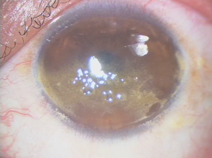
Zhang B, Yao Y. Gelatinous drop-like corneal dystrophy with a novel mutation of TACSTD2 manifested in combination with spheroidal degeneration in a Chinese patient. Molecular Vision. 2010 Aug 11; 16:1570. Figure 1. PMCID: PMC2927379. License: CC BY.
Anterior segment photograph showing clusters of amber-colored spherical deposits in the superficial cornea and anterior stroma. It shows shiny deposit nodules predominantly in the exposed area.
Deposit characteristics: Yellow-white to golden or transparent raised lesions. Similar in extent and shape to band keratopathy, but the deposit characteristics differ.
Distribution: Begins at the peripheral cornea at the 3 and 9 o’clock positions and progresses centrally. Commonly occurs in the interpalpebral fissure.
Advanced cases: Present as plaque-like lesions crossing the central cornea. Deposits may become nodular.
Superficial vascular invasion: May be accompanied by superficial vascular invasion.
Anterior segment OCT (AS-OCT) findings: Depicts subepithelial fibrosis and anterior stromal scarring. In Grade 3 cases, stromal scarring at a depth of approximately 60 μm was confirmed1).
Ultraviolet (UV) exposure is considered the most important environmental factor. It is associated with elastoid degeneration seen in pterygium and pinguecula. It is presumed that UV interaction causes serum proteins from the limbal vessels to diffuse into the cornea, leading to degeneration and accumulation.
UV exposure: The greatest risk factor. UV reflected from ice, snow, and desert areas contributes.
Climatic conditions: High temperature, low humidity, sandstorms, and low rainfall increase risk. Prevalence is high in southwestern Rajasthan, India2).
Occupational exposure: Burns from welding are also recognized as an occupational risk.
Individual Factors and Ocular Diseases
Aging: Incidence increases with age.
Pre-existing ocular diseases: Keratitis, lattice corneal dystrophy, glaucoma, etc., are risk factors for secondary corneal type.
Others: Dry eye, malnutrition, corneal trauma, and microtrauma from wind, sand, and ice are involved.
QWhy is spheroidal degeneration more common in certain regions?
A
Because UV exposure is the greatest risk factor, the prevalence is high in high-latitude areas where UV reflects from snow and ice (e.g., Labrador region) and in low-latitude areas near the equator where UV is strong in desert regions. In southwestern Rajasthan, India, the prevalence of spheroidal degeneration is particularly high due to high temperatures, sandstorms, and low rainfall 2).
Slit-lamp microscopy: Yellow-brown spherical deposits are observed under the corneal epithelium in the interpalpebral fissure. They appear droplet-like at low magnification but disappear at high magnification.
Johnson-Ghosh grading: The extent of the lesion and its impact on vision are evaluated from Trace to Grade 4.
Imaging Tests
Anterior segment OCT: Objectively evaluates the depth and extent of deposits. It can measure the depth of subepithelial fibrosis and anterior stromal scarring 1).
Histological examination: Stains eosinophilic with H&E and blue-green with toluidine blue. Not essential for diagnosis.
Band keratopathy: Similar in extent and shape, but differs in that it involves calcium salt deposits and appears white and opaque.
Gelatinous drop-like corneal dystrophy: Bilateral, onset in childhood. Amyloid deposits progress from the central cornea to the periphery. Caused by abnormalities in the TACSTD2 gene.
Salzmann nodular degeneration: Nodular degeneration of the corneal stroma, differing in distribution and the nature of deposited material.
Vogt limbal girdle: Calcium salt deposits in the peripheral cornea, requiring differentiation as an age-related change.
Superficial keratectomy: Performed when there is decreased vision or foreign body sensation. Deposits are mechanically removed.
PTK (excimer laser therapeutic keratectomy): Used to remove superficial deposits.
Superficial corneal transplantation: Performed in cases where superficial keratectomy is insufficient.
Deep anterior lamellar keratoplasty (DALK): Indicated for cases with deep stromal scarring. There is a report of a patient with Grade 3 spheroidal degeneration who underwent DALK and achieved 6/9 vision one month postoperatively1). However, the cornea in spheroidal degeneration is hard, and big bubble formation may be difficult1).
In cataracts complicated by spheroidal degeneration, intraoperative visibility is reduced due to corneal opacity2). In areas where penetrating keratoplasty is difficult to perform, SICS (small incision cataract surgery) + IOL implantation is an effective option, with visual improvement achieved in 92.85% of patients2).
In a study of 56 cases of spheroidal degeneration with cataract in a tribal population in Rajasthan, SICS improved mean best corrected visual acuity (BCVA) from 2.3±0.67 logMAR preoperatively to 0.4±0.22 logMAR at one month postoperatively (P < 0.0001). 63.3% achieved vision of 6/6 to 6/122).
QIs corneal transplantation effective?
A
Superficial keratectomy and deep anterior lamellar keratoplasty (DALK) are effective for spheroidal degeneration. In Grade 3 cases, DALK has been reported to achieve visual acuity of 6/9 postoperatively 1). However, the cornea in spheroidal degeneration is hard, making big bubble formation difficult 1). Additionally, recurrence is possible after superficial keratectomy or superficial keratoplasty.
Pathologically, hyaline-like material accumulates in the subepithelium and Bowman’s layer. The deposited material is composed of proteins. It is thought to be granular protein secreted by keratocytes that deposits on adjacent collagen fibers.
Several hypotheses exist regarding the origin of the deposited material. A leading hypothesis proposes that plasma proteins (immunoglobulins and albumin) diffuse into the cornea from the limbal circulation and are denatured and accumulate due to ultraviolet radiation. Another hypothesis suggests that secretory products from the stroma or epithelium (incomplete collagen or degraded stromal collagen) deposit.
It is associated with elastoid degeneration seen in pterygium and pinguecula. Chronic tissue damage from ultraviolet radiation is considered a common pathological basis.
Under light microscopy, extracellular deposits of various sizes, spherical or elongated, are observed in the subepithelium, Bowman’s layer, and superficial stroma. In advanced stages, Bowman’s layer is focally disrupted or absent. Inflammatory cells are usually absent. Electron microscopy shows fine granular structures accumulating on collagen bundles.
QWhat is the identity of the deposited material?
A
The deposited material is composed of proteins. It is thought to be granular protein secreted by keratocytes that deposits on collagen fibers. A leading hypothesis is that plasma proteins (immunoglobulins and albumin) diffuse from limbal vessels and are denatured and accumulate due to ultraviolet radiation. It stains eosinophilic with H&E and blue-green with toluidine blue.
The efficacy of deep anterior lamellar keratoplasty for spheroidal degeneration has been reported. In cases of Grade 3 spheroidal degeneration treated with DALK, big bubble formation was difficult, so the procedure was converted to manual dissection 1). Because the cornea in spheroidal degeneration is hard, after big bubble formation, the host Descemet’s membrane bulged into the anterior chamber, causing secondary angle-closure glaucoma due to reverse pupillary block1).
In a case of deep anterior lamellar keratoplasty for Grade 3 central spheroidal degeneration in a 65-year-old male, acute angle-closure glaucoma with intraocular pressure of 46 mmHg occurred on postoperative day 1. Anterior segment OCT revealed bulging of the host Descemet’s membrane and residual stroma. Angle closure was relieved by decompression of the big bubble, and visual acuity of 6/9 was achieved at 1 month postoperatively1).
In regions with high prevalence of spheroidal degeneration, the safety and efficacy of SICS for complicated cataracts have been investigated2). Among 124 patients with hard cataracts and ocular complications, 45.16% had concurrent spheroidal degeneration2). Although reduced intraoperative visibility due to corneal opacity leads to difficulty in capsulorhexis, no serious complications were observed2).
Kodavoor SK, Ravi P. Secondary angle closure glaucoma due to iatrogenic big bubble formation in manual DALK for stromal degeneration: An unusual presentation. Romanian journal of ophthalmology. 2023;67(3):309-311. doi:10.22336/rjo.2023.50. PMID:37876517; PMCID:PMC10591435.
Gajraj M, Mohan A. Safety and efficacy of manual small-incision cataract surgery in patients with brunescent and black cataracts and other ocular comorbidities. Indian J Ophthalmol. 2022;70(11):3898-3903.