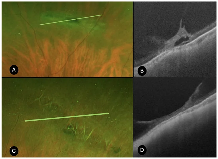
Bacherini D, et al. Characterization of Peripheral Retinal Degenerations and Rhegmatogenous Lesions Using Ultra-Widefield Swept Source OCT Integrated with a Novel Scanning Laser Ophthalmoscope. Diagnostics (Basel). 2025. Figure 1. PMCID: PMC12650825. License: CC BY.
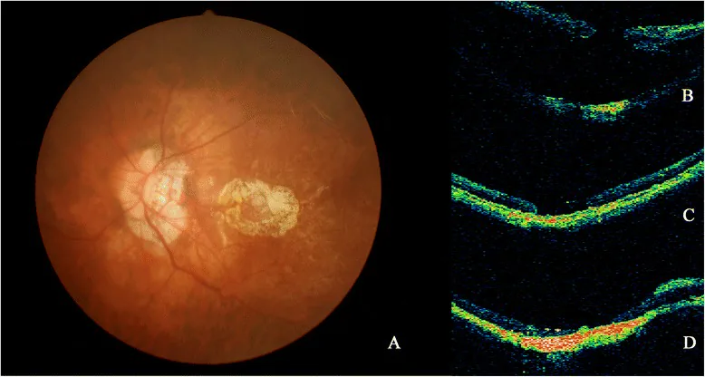
Nan Hong; Bai-shuang Huang; Jian-ping Tong. Primary silicone oil tamponade and internal limiting membrane peeling for retinal detachment due to macular hole in highly myopic eyes with chorioretinal atrophy. BMC Ophthalmol. 2015 Nov 11; 15:165 Figure 2. PMCID: PMC4642637. License: CC BY.
Fundus photograph and OCT images (a, b, c and d) from Patient 20. a PCA around MH was shown on fundus photograph. b RD caused by MH developed. c After the SO removal, retinal reattachment was achieved, MH was still open on OCT. d After the second silicone oil removal, the patient achieved retina reattachment despite persistent open of the MH
鑑別診断として類嚢胞変性・敷石状変性・white without pressureを念頭に置く。敷石状変性は脈絡膜毛細血管の循環不全による変性であり、硝子体液化や円孔・裂孔の原因にはならない。white without pressureは強膜圧迫なしに白色調変化を呈する所見であり、格子状変性との鑑別が必要な場合がある。類嚢胞変性は内顆粒層の空洞化であり、円孔形成とは別の機序である。
Kim SJ, Bailey ST, Kovach JL, Lim JI, Vemulakonda GA, Ying GS, et al. Posterior Vitreous Detachment, Retinal Breaks, and Lattice Degeneration Preferred Practice Pattern®. Ophthalmology. 2025;132(4):P163-P196. doi:10.1016/j.ophtha.2024.12.023. PMID:39918519.
Maltsev DS, Kulikov AN, Shaimova VA, Burnasheva MA, Vasiliev AS. Spotlight on Lattice Degeneration Imaging Techniques. Clinical ophthalmology (Auckland, N.Z.). 2023;17:2383-2395. doi:10.2147/OPTH.S405200. PMID:37605766; PMCID:PMC10440085.
Eaddy IC, Moushmoush O, Sabbagh O, Barazi MD, Sabbagh O.. Horseshoe Retinal Tear Minutes After Use of a New Pilocarpine Formulation in a Presbyopic, Emmetropic Man. J Vitreoretin Dis. 2025;9(1):105-108. doi:10.1177/24741264241255589. PMID:39554627; PMCID:PMC11562214.
Manoli K, Ching J. A macular horseshoe tear following posterior vitreous detachment and longstanding branch retinal vein occlusion. GMS Ophthalmol Cases. 2025;15:Doc16. doi:10.3205/oc000264.
Sasongko MB, Wan R, Ho IV. Large, star-shaped retinal tear associated with orbital cosmetic filler. American journal of ophthalmology case reports. 2022;25:101342. doi:10.1016/j.ajoc.2022.101342. PMID:35243133; PMCID:PMC8859732.
Byer NE. Long-term natural history of lattice degeneration of the retina. Ophthalmology. 1989;96(9):1396-401; discussion 1401-2. doi:10.1016/s0161-6420(89)32713-8. PMID:2780007.
Haimann MH, Burton TC, Brown CK. Epidemiology of retinal detachment. Archives of ophthalmology (Chicago, Ill. : 1960). 1982;100(2):289-92. doi:10.1001/archopht.1982.01030030291012. PMID:7065947.
Byer NE. Subclinical retinal detachment resulting from asymptomatic retinal breaks: prognosis for progression and regression. Ophthalmology. 2001;108(8):1499-1503; discussion 1503-1504. PMID: 11470709. doi:10.1016/S0161-6420(01)00652-2.
The Eye Disease Case-Control Study Group. Risk factors for idiopathic rhegmatogenous retinal detachment. Am J Epidemiol. 1993;137(7):749-757. doi:10.1093/oxfordjournals.aje.a116735.
N Byer. What happens to untreated asymptomatic retinal breaks, and are they affected by posterior vitreous detachment?. Ophthalmology. 1998;105(6):1045-1050. doi:10.1016/s0161-6420(98)96006-7.
Morano MJ, Khan MA, Zhang Q, Halfpenny CP, Wisner DM, Sharpe J, Li A, Tomaiuolo M, et al. Incidence and Risk Factors for Retinal Detachment and Retinal Tear after Cataract Surgery: IRIS® Registry (Intelligent Research in Sight) Analysis. Ophthalmology science. 2023;3(4):100314. doi:10.1016/j.xops.2023.100314. PMID:37274012; PMCID:PMC10239011.
Coffee RE, Westfall AC, Davis GH, Mieler WF, Holz ER.. Symptomatic posterior vitreous detachment and the incidence of delayed retinal breaks: case series and meta-analysis. Am J Ophthalmol. 2007;144(3):409-413. doi:10.1016/j.ajo.2007.05.002. PMID:17583667.
Michael I. Seider, Carol Conell, Ronald B. Melles. Complications of Acute Posterior Vitreous Detachment. Ophthalmology. 2022;129(1):67-72. doi:10.1016/j.ophtha.2021.07.020.
Wilkinson CP. Interventions for asymptomatic retinal breaks and lattice degeneration for preventing retinal detachment. The Cochrane database of systematic reviews. 2014;2014(9):CD003170. doi:10.1002/14651858.CD003170.pub4. PMID:25191970; PMCID:PMC4423540.
Johnson MW. Posterior vitreous detachment: evolution and role in macular disease. Retina. 2012;32 Suppl 2:S174-S178. doi:10.1097/IAE.0b013e31825bef62.
Curran CD, Adams OE, Vagaggini T, Sodhi GS, Prairie ML, Baker MJ, Sastry A, Ryan EH, Parke DW, Mittra RA, Dev S, Tang PH.. PROPHYLACTIC TREATMENT OF LATTICE DEGENERATION IN FELLOW EYES AFTER REPAIR OF UNCOMPLICATED PRIMARY RHEGMATOGENOUS RETINAL DETACHMENT. Retina. 2024;44(1):63-70. doi:10.1097/iae.0000000000003908. PMID:37536462.