Disease Overview
Official name: RAMSVPS syndrome
Former name: FRAM
Inheritance pattern: Autosomal recessive
Causative gene: IGFBP7 (c.830-1G>A)

Familial Retinal Arterial Macroaneurysms (FRAM) is a rare autosomal recessive disease caused by mutations in the IGFBP7 gene. It was first reported in 2002 by Dhindsa & Abboud. 1)
Subsequently, after identification of IGFBP7 mutations and confirmation of supravalvular pulmonic stenosis by echocardiography, it is now renamed as RAMSVPS (Retinal Arterial Macroaneurysms with Supravalvular Pulmonic Stenosis) syndrome. 1)
This disease is characterized by progressive bilateral multiple retinal arterial macroaneurysms. The causative mutation is a splicing mutation c.830-1G>A (homozygous) in IGFBP7 (4q12), leading to upregulation of the BRAF/MEK/ERK pathway. 1)
Reported cases are limited to 15 families and 30 individuals, mainly from Saudi Arabia and the UAE. 1) The mean age at initial diagnosis is 12.5 years (range 1–34 years), with no sex predilection.
Disease Overview
Official name: RAMSVPS syndrome
Former name: FRAM
Inheritance pattern: Autosomal recessive
Causative gene: IGFBP7 (c.830-1G>A)
Epidemiology
Number of reported patients: 30 (15 families)
Mean age at first visit: 12.5 years (range 1–34 years)
Sex predilection: None
Ethnicity: Arabian Peninsula origin
They are the same disease. First reported as FRAM in 2002, an IGFBP7 mutation was identified in 2011, and after echocardiography confirmed supravalvular pulmonary stenosis, it was renamed RAMSVPS. 1)
The main complaint is decreased visual acuity due to hemorrhage and exudation from the macroaneurysm into the macula. In reported cases, the best-corrected visual acuity (BCVA) at initial visit was 20/300 in the right eye and 20/60 in the left eye. 1)
Some asymptomatic patients with a family history are detected through screening examinations.
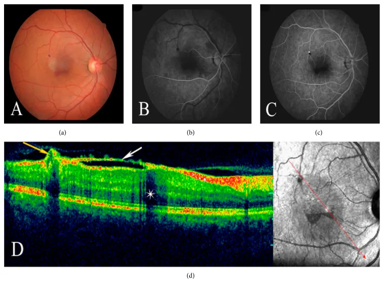
Arterial beading and macroaneurysms are the most characteristic findings, observed in all patients (100%). 1) Persistent hyaloid artery is present in 88–90% of cases. 1) Recurrent hemorrhage/exudation (vitreous/subretinal) occurs in 71%, and submacular gliosis in 35%. 1)
Macroaneurysms with leakage are clearly visualized on fluorescein angiography (FA). Peripheral telangiectasia, dropout, and arterial beading are also observed simultaneously. 1)
Characteristic Ocular Findings
Arterial beading + macroaneurysms: 100%
Persistent hyaloid artery: 88–90%
Recurrent hemorrhage/exudation: 71%
Other Ocular Findings
Retinal artery narrowing/sheathing: 42%
Coats-like findings: 42%
Submacular gliosis: 35%
In severe cases, macroaneurysms with leakage lead to intraretinal/subretinal exudates and progress to exudative retinal detachment. Coats-like findings (peripheral telangiectasia/nonperfusion) are observed in 42%. 1)
The causative gene is IGFBP7 (4q12), and homozygosity for the splicing mutation c.830-1G>A has been confirmed in all reported cases. 1) The inheritance pattern is autosomal recessive, and it is frequently observed in families with consanguineous marriage. 1)
IGFBP7, also called angiomodulin, is a vascular secretory factor. 1) In the retina, it is specifically expressed in the endothelium of large smooth muscle vessels, but not in capillaries. 1) This expression pattern is thought to explain why beading and macroaneurysms are confined to the major arterial trunks.
Histologically, multiple linear breaks are observed in the retinal artery wall. The frequencies of systemic complications are shown below.
| Systemic complication | Frequency | Outcome |
|---|---|---|
| Pulmonary artery stenosis | 65% | 47% surgery, 13% death |
| Supravalvular aortic stenosis | 22% | — |
| Cerebrovascular lesions | 3 cases | Stroke/hemorrhagic death |
It is an autosomal recessive disorder, and definitive diagnosis is possible by identifying IGFBP7 c.830-1G>A. 1) It is more common in families with consanguineous marriage, and carriers (heterozygotes) are asymptomatic.
Arterial beading and macroaneurysms of the major arterial trunks are observed in all patients (100%). 1) FA reveals macroaneurysms with leakage, peripheral telangiectasia, and dropout. 1)
Identification of IGFBP7 c.830-1G>A is essential for definitive diagnosis. This mutation was not found in 300 healthy controls. 1)
Echocardiographic screening for pulmonary artery stenosis is essential (65% have cardiac disease). 1) For detection of coronary artery aneurysms and systemic arterial stenosis, systemic vascular evaluation including cardiac catheterization is performed. 1)
FRAM must be differentiated from diseases with similar fundus findings.
| Disease Name | Main Features | Points of Differentiation from FRAM |
|---|---|---|
| Coats disease | Unilateral, idiopathic | Telangiectasia is predominant |
| IRVAN | Non-familial, inflammatory | Vasculitis, anterior chamber cells present |
| Takayasu arteritis | Large vessel vasculitis | Differentiated by genetic testing |
FRAM is bilateral and familial, characterized by beading of major arterial trunks and aortic aneurysms. Coats disease is unilateral and idiopathic, primarily involving telangiectasia. IRVAN is non-familial and associated with vasculitis and anterior chamber inflammation. Persistent hyaloid artery (88–90%) is also an important distinguishing feature of FRAM. 1)
For leaking macroaneurysms, retinal grid laser photocoagulation is performed to stop lipid exudation. Multiple sessions may be required. 1)
In reported cases, multiple laser photocoagulation sessions over two years in the right eye resulted in cessation of leakage and regression of exudates. 1)
Note that after spontaneous rupture of a macroaneurysm, the aneurysm becomes naturally occluded and fibrotic, so laser treatment is unnecessary. 1)
Panretinal photocoagulation is performed for ischemic areas. 1)
Performed in cases with vitreous hemorrhage or submacular hemorrhage. 1) In reported cases, a combination of cataract surgery, vitrectomy, membrane peeling, and SF6 gas tamponade improved BCVA to 20/80. 1)
Reports exist for bevacizumab and others, but their role in RAMSVPS is not established. 1)
Cardiovascular evaluation and management are essential; coronary artery bypass surgery has been performed in some cases. 1) Collaboration with cardiology affects life prognosis.
It varies by case. Laser photocoagulation for leaking macroaneurysms can control exudation, but if repeated bleeding and exudation cause submacular gliosis, visual recovery becomes difficult. 1) Systemic management (cardiovascular) is also important as it directly affects life prognosis.
IGFBP7 mutations impair normal IGFBP7 production. 1) IGFBP7 (angiomodulin) is a CNS vessel-specific angiocrine factor that blocks VEGF-induced angiogenesis when VEGF-A signaling drives brain angiogenesis. 1)
Experiments in zebrafish show that blocking angiomodulin leads to defects in vessel sprouting and patterning. 1)
In the retina, IGFBP7 is specifically expressed in the endothelium of large smooth muscle vessels and is not expressed in capillaries. 1) This expression distribution is the pathological basis for beading and aneurysms occurring only in the main arterial trunks. Aortic aneurysms form at sites of reduced mechanical integrity of the vessel wall where pressure is highest. 1)
Three parallels are observed between retinal findings and systemic arterial findings. 1)
Regarding the pathogenesis of Coats disease-like findings, disruption of the inner blood-retinal barrier (iBRB) due to pericyte loss is thought to cause abnormal capillary dilation. 2)
Reports of RAMSVPS cases with coronary artery aneurysms are revealing that systemic vascular lesions are more extensive than previously recognized. 1) Additionally, the similarity of arterial findings between Takayasu arteritis and RAMSVPS may suggest the existence of an undiscovered common pathological basis. 1)
All reported cases to date are limited to individuals from the Arabian Peninsula, but there may be undiagnosed or misdiagnosed cases outside the Arabian Peninsula. It has been suggested that some cases diagnosed as Coats disease or IRVAN may include FRAM. 1)
The efficacy of anti-VEGF drugs for RAMSVPS has not been established, and further investigation is awaited. 1)
Retinal macroaneurysm formation secondary to retinal vein occlusion has also been reported, 3) and pathophysiological studies of FRAM may provide clues to elucidate the mechanism of aneurysm formation.