
Binocular Vision Testing
Key points at a glance
Section titled “Key points at a glance”1. What Is Binocular Vision Testing?
Section titled “1. What Is Binocular Vision Testing?”Binocular vision is defined as the sensation in which the visual input from the right eye and the left eye is recognized simultaneously in the brain’s visual centers. Binocular vision testing objectively evaluates this function.
Three Elements of Binocular Vision Function
Section titled “Three Elements of Binocular Vision Function”Binocular vision function consists of the following three elements and has a stepwise hierarchical structure.
- Simultaneous perception (simultaneous perception): The ability to recognize the visual input from both eyes at the same time. The first stage, forming the foundation of binocular vision function
- Fusion (fusion): The ability to superimpose similar images projected to both eyes and recognize them as one image (sensory fusion). The second stage, obtained only after simultaneous perception is established
- Stereopsis (stereopsis): The highest-level function in which the brain detects binocular disparity and converts it into depth. The third stage, acquired only after simultaneous perception and fusion are established
Stereopsis is the highest level of binocular vision and is responsible for depth perception and spatial awareness in daily life.
The three elements are simultaneous perception, fusion, and stereopsis, forming a stepwise hierarchy in that order. Simultaneous perception is the basic function of recognizing visual input from both eyes at the same time; fusion is the function of combining the images seen by the two eyes into one; stereopsis is the highest-level function of detecting depth from binocular disparity. Stereopsis can be obtained only when both simultaneous perception and fusion are established. In strabismus and amblyopia, the higher levels of this hierarchy are affected first, so evaluating all three elements is important when setting treatment goals.
Conditions for binocular vision to develop
Section titled “Conditions for binocular vision to develop”For binocular vision to develop normally, all of the following five conditions must be met.
- Binocular vision cells are present in the visual center (cerebrum)
- Both eyes have good visual acuity
- There is no aniseikonia (there is not a large difference in the size of the image formed on the retina between the two eyes)
- There is no strabismus
- There is normal retinal correspondence
If any of these conditions is impaired, some or all of binocular vision will be affected. Strabismus, amblyopia, and anisometropia are the main causes of binocular vision problems.
Indications for binocular vision testing
Section titled “Indications for binocular vision testing”The main indications for binocular vision testing are as follows.
- Preoperative and postoperative evaluation of strabismus (confirmation of recovery of binocular vision)
- Confirmation of treatment effect in amblyopia (assessment of occlusion therapy and patching of the sound eye)
- Evaluation of phoria (confirmation of fusion ability in small phoria)
- Determination of indication for fusion training and monitoring of treatment effect
- Vision screening in children (such as preschool examinations)
- Evaluation of retinal correspondence and simultaneous perception in patients who complain of diplopia
2. Worth 4-light Test
Section titled “2. Worth 4-light Test”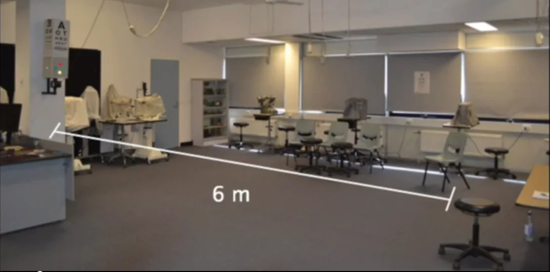
The Worth four-light test is a representative binocular vision test that uses red-green complementary filters to separate the two eyes and evaluates simultaneous perception, fusion, and retinal correspondence12.
Principle
Section titled “Principle”Red and green are complementary colors. Through a red filter, the green target cannot be seen, and through a green filter, the red target cannot be seen. Using this property, each eye is separated by a red filter (right eye) and a green filter (left eye), and the four lights (top: red, left and right: green, bottom: white) are shown; the response is used to determine simultaneous perception, fusion, suppression, and diplopia.
Interpretation
Section titled “Interpretation”| How it appears | Number of lights | Interpretation |
|---|---|---|
| Two red lights + two green lights (the white light appears red or green) are seen correctly | 4 lights | Normal, fusion present |
| Only two red lights are seen | 2 lights (red) | Suppression of the left eye (green-filter side) |
| Only the two green lights are visible | 2 lights (green) | Suppression of the right eye (the red-filter side) |
| Two columns of lights are visible | 5 lights | Diplopia (no simultaneous perception, no fusion) |
Practical considerations
Section titled “Practical considerations”- It is preferable to perform the test at both near (33 cm) and distance (3–6 m). If the results differ between near and distance, this may reflect distance-dependent ocular deviation or a difference in fusion ability
- When judging retinal correspondence, it is important to know the eye position in advance (such as by cover test)
- In children, Berens 3-light test is used, reducing the number of lights to three based on the same principle
- To quantitatively assess the size and depth of the suppression zone, expanded testing methods using tablet devices (such as W4DApp) have also been developed in recent years1
- It has been reported that even in patients with red-green color vision deficiency, the Worth 4-dot test is useful for assessing binocular vision function2
If five lights are seen, it indicates diplopia, meaning there is no simultaneous vision and no fusion, or they are not functioning. The images from the right and left eyes are not combined into one, so they are seen as two rows. The upper two red lights and the lower two green lights are seen separately without overlapping. This may be seen in intermittent strabismus, constant exotropia, or sensory fusion disorder. It should be evaluated after confirming eye alignment and combined with other binocular vision tests.
3. Bagolini striated lens test
Section titled “3. Bagolini striated lens test”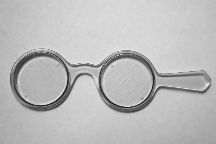
The Bagolini striated glass test is a test that can assess binocular vision function in a state closest to everyday vision3.
Principle and procedure
Section titled “Principle and procedure”Bagolini striated glasses are a pair of plane lenses (glass that is almost optically neutral) with fine parallel scratches. When you wear these lenses and look at a point light source such as a penlight, a streak of light appears from the light source in a direction perpendicular to the scratches on the lens. The right-eye and left-eye lenses are arranged so that the directions of the streaks are at right angles, and in normal binocular vision two streaks are seen crossing at right angles around the penlight.
Interpretation
Section titled “Interpretation”- Two perpendicular streaks are seen centered on the light source → normal correspondence, fusion present (normal binocular vision)
- Only one streak is seen → suppression present (information from one eye is being suppressed)
- If the two stripe images are misaligned (the intersection does not match the penlight position) → diplopia (diplopia due to anomalous retinal correspondence or strabismus)
Characteristics and clinical significance
Section titled “Characteristics and clinical significance”The greatest feature of the Bagolini striated lens test is that it can be performed in a natural state that is closest to everyday vision. Because binocular separation is weak, suppression tends to occur easily (the suppression mechanism works as it does in natural viewing), and it is excellent for detecting abnormal correspondence (anomalous retinal correspondence) 4. It has been shown from neuroanatomical studies and analysis of clinical cases that anomalous retinal correspondence is likely to occur in small-angle strabismus (4–5 degrees = 8–10 prism diopters) 4.
On the other hand, the sensitivity for detecting suppression is lower than with other tests (Worth 4-dot test and major amblyoscope), so note that mild suppression is difficult to detect. A three-light modified method based on Bagolini striated glasses (the starlight test) is also used for binocular visual field screening 3.
4. Afterimage test (Bielschowsky afterimage test)
Section titled “4. Afterimage test (Bielschowsky afterimage test)”The afterimage test (Bielschowsky afterimage test) is a test that independently forms afterimages on each eye’s fovea and assesses retinal correspondence, simultaneous perception, and suppression from their spatial relationship.
Principle and procedure
Section titled “Principle and procedure”If central fixation is maintained, retinal correspondence and simultaneous perception can be assessed regardless of eye position.
The procedure is as follows.
- Cover the strabismic eye and shine a horizontal flash of light (horizontal line) on the fixing eye (dominant eye)
- Cover the fixing eye and shine a vertical flash of light (vertical line) on the strabismic eye
- With both eyes open, ask the examinee how the afterimage appears
Interpretation
Section titled “Interpretation”| How the afterimage appears | Interpretation |
|---|---|
| The vertical and horizontal afterimages are orthogonal at the center (crossing in a cross shape) | Normal correspondence (positive Hering-Bielschowsky test) |
| The vertical and horizontal afterimages are separated (do not cross) | Abnormal correspondence (abnormal retinal correspondence) |
| Only one afterimage is seen | Suppression (the fovea of the non-fixating eye is suppressed) |
Clinical significance and points to note
Section titled “Clinical significance and points to note”The afterimage test provides the strongest degree of binocular dissociation and has the highest sensitivity for detecting suppression. It is used to check for abnormal correspondence when determining indications for strabismus surgery and in postoperative assessment.
However, because central fixation is a prerequisite, it cannot be assessed in cases with eccentric fixation (a state in which central fixation has been lost).
5. Comparison and selection of each test method
Section titled “5. Comparison and selection of each test method”The various binocular vision tests each have different characteristics, so it is important to choose the appropriate one according to the testing purpose and clinical situation.
Comparison of the characteristics of each test
Section titled “Comparison of the characteristics of each test”| Test | Closeness to everyday viewing | Suppression detection | Detection of abnormal correspondence | Ease of fusion |
|---|---|---|---|---|
| Bagolini striated lenses | Closest | Hard to detect | Easily detected | Fusion is easy |
| Worth 4-dot test | Moderate | Moderate | Moderate | Moderate |
| Large synoptophore (synoptophore) | Far | Easy to detect | Hard to elicit | Difficult to elicit |
| Afterimage test | Farthest | Easiest to detect | Hard to elicit | Most difficult to elicit |
How to Choose
Section titled “How to Choose”The stronger the binocular dissociation of a test, the more likely it is to detect deep (true) anomalous retinal correspondence; the closer a test is to everyday vision, the better it can assess how vision actually appears.
- If you want to understand real-life performance → Bagolini striated lens
- If you want to reliably check for suppression or double vision → Worth 4-dot test / synoptophore
- Final confirmation of anomalous retinal correspondence / preoperative assessment → afterimage test
- Pediatric screening / quick check → Worth 4-dot test (Behrens 3-light test)
These tests should not be used alone; the rule is to combine several and assess the patient comprehensively.
The Bagolini striated lens test can assess binocular function in a state closest to everyday vision. This lens is made by putting fine scratches on an almost optically neutral flat piece of glass, so wearing it does not greatly change the visual field or visual acuity. For that reason, it can assess retinal correspondence and fusion under conditions close to ordinary binocular fusion, and it is excellent for detecting anomalous retinal correspondence. On the other hand, because binocular dissociation is weak, mild suppression may be difficult to detect.
6. Development of Binocular Vision and Clinical Significance
Section titled “6. Development of Binocular Vision and Clinical Significance”Developmental Timeline
Section titled “Developmental Timeline”Binocular visual function develops rapidly from early infancy. Studies using VEP (visual evoked potentials) have confirmed the following developmental pattern.
- By 2 months after birth: binocular vision appears
- At 3 to 5 months after birth: fusion begins
- By 20 weeks after birth: stereopsis is detected in more than 75% of children5
- At 6 to 7 months after birth: the stereoacuity threshold reaches nearly adult levels5
If strabismus, amblyopia, or anisometropia occurs during this period, the normal development of binocular visual function is hindered. With appropriate treatment during the sensitive period (critical period), recovery of binocular visual function can be expected.
Relation to amblyopia and strabismus treatment
Section titled “Relation to amblyopia and strabismus treatment”The final goal of amblyopia treatment is not only improvement of visual acuity, but also recovery of binocular visual function.
- Sound-eye occlusion therapy (patching): this promotes visual improvement in the amblyopic eye, but binocular vision does not function during occlusion, so after treatment ends, binocular vision testing is used to confirm the state of fusion and stereopsis
- Preoperative evaluation before strabismus surgery: preoperative binocular vision tests (such as the Worth 4-dot test and afterimage test) are used to assess anomalous correspondence and suppression, and to judge the possibility of postoperative recovery of binocular visual function
- Postoperative evaluation after strabismus surgery: postoperative binocular vision tests are used to confirm the recovery of simultaneous perception, fusion, and stereopsis after eye position correction
- Management of phoria: in small phoria, fusional ability is often preserved, but symptoms may appear if fusional ability declines due to fatigue or aging, so quantitative assessment of fusional ability is useful
How it is used compared with stereopsis testing
Section titled “How it is used compared with stereopsis testing”The Worth 4 Dot test, Bagolini striated lenses, and the afterimage test are mainly used to evaluate simultaneous perception, fusion, retinal correspondence, and suppression. In clinical practice, when simultaneous perception and fusion are confirmed with these tests, stereopsis testing (Titmus fly test, Lang stereotest, TNO test, etc.) is then used to measure whether stereopsis is present and how deep it is.
Stereopsis depends on both the sensory and motor aspects of binocular vision and is clinically useful as a highly sensitive indicator of whether strabismus is present and how severe it is6. For a detailed evaluation of stereopsis, see the Stereopsis Testing section.
7. References
Section titled “7. References”Footnotes
Section titled “Footnotes”-
Webber AL, Mandall TR, Molloy DT, Lister LJ, Birch EE. Worth 4 Dot App for Determining Size and Depth of Suppression. Transl Vis Sci Technol. 2020;9(2):3. PMID: 32818097. ↩ ↩2
-
Bak E, Yang HK, Hwang JM. Validity of the Worth 4 Dot Test in Patients with Red-Green Color Vision Defect. Optom Vis Sci. 2017;94(5):626-629. PMID: 28234793. ↩ ↩2
-
Hirai T, Arai M, Ito Y, Sato M. Modified Bagolini striated glass test: clinical applications of starlight test in binocular visual field screening. Br J Ophthalmol. 1998;82(11):1288-1293. PMID: 9924335. ↩ ↩2
-
Wong AM, Lueder GT, Burkhalter A, Tychsen L. Anomalous retinal correspondence: neuroanatomic mechanism in strabismic monkeys and clinical findings in strabismic children. J AAPOS. 2000;4(3):168-174. PMID: 10849394. ↩ ↩2
-
Birch E, Petrig B. FPL and VEP measures of fusion, stereopsis and stereoacuity in normal infants. Vision Res. 1996;36(9):1321-1327. PMID: 8711910. ↩ ↩2
-
Read JC. Stereo vision and strabismus. Eye (Lond). 2015;29(2):214-224. PMID: 25475234. ↩