
Fingolimod-associated macular edema
Key Points at a Glance
Section titled “Key Points at a Glance”1. What is Fingolimod-Associated Macular Edema?
Section titled “1. What is Fingolimod-Associated Macular Edema?”Fingolimod (Imusera/Gilenya) is the first oral disease-modifying therapy (DMT) for relapsing-remitting multiple sclerosis (RRMS). It modulates the sphingosine-1-phosphate (S1P) receptor and suppresses autoimmune demyelination in the central nervous system by blocking the release of lymphocytes from lymph nodes1).
Macular edema (ME) is a serious ocular adverse effect of this drug, known as fingolimod-associated macular edema (FAME)2). It was launched in Japan in 2011, and due to limited experience with long-term administration, a full-case survey is mandated as a condition of approval. In overseas clinical trials, the incidence of macular edema was 0.2% at 0.5 mg/day and 1.4% at 1.25 mg/day. In domestic clinical trials, no macular edema was observed.
Newer S1P receptor modulators of the same class (such as siponimod) have not reported similar risks of macular edema in clinical trials.
The incidence at the approved dose (0.5 mg/day) is low, ranging from 0% to 0.5% in overseas clinical trials. At a higher dose (1.25 mg/day), it increases to 1% to 1.6%. No cases were reported in domestic clinical trials. For details, see the “Causes and Risk Factors” section.
2. Main Symptoms and Clinical Findings
Section titled “2. Main Symptoms and Clinical Findings”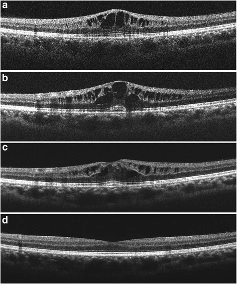
Subjective Symptoms
Section titled “Subjective Symptoms”The main symptoms reported by FAME patients are as follows4).
- Decreased central vision/blurring: This is the most common complaint.
- Color vision changes: Patients notice abnormalities in how colors appear.
- Metamorphopsia: Objects appear distorted.
- Micropsia: Objects appear smaller.
- Scotoma: Relative or absolute scotomas are observed.
Most cases of macular edema observed in overseas clinical trials were asymptomatic. It may be first discovered during routine examinations 3 to 4 months after starting treatment.
Clinical Findings (Findings Confirmed by Physician Examination)
Section titled “Clinical Findings (Findings Confirmed by Physician Examination)”- Decreased visual acuity: Measured using a Snellen chart, etc.
- Decreased contrast sensitivity: Assessed using a Pelli-Robson chart, etc.
- Color vision abnormalities: Detected by color vision testing.
- Metamorphopsia: Confirmed using an Amsler grid.
- Dilated fundus examination findings: Retinal thickening, intraretinal cysts, and changes in macular color. May be more clearly visible with green light.
Moderate to severe ME (foveal thickness >300 μm) may be detectable by slit-lamp microscopy4). Mild ME (foveal thickness 201–300 μm) requires OCT for detection4).
3. Causes and Risk Factors
Section titled “3. Causes and Risk Factors”The development of FAME is influenced by fingolimod dose and duration, patient age, and comorbidities.
The incidence of macular edema in major clinical trials is shown below.
| Trial/Population | 0.5 mg group | High-dose group |
|---|---|---|
| FREEDOMS/TRANSFORMS trials (MS) | 0–0.5% | 1–1.6% (1.25 mg) |
| Renal transplant trials (+ cyclosporine) | — | 1.3% (2.5 mg) / 2.2% (5.0 mg) |
The incidence is dose-dependent and is low at the approved dose (0.5 mg/day)9, 10, 11).
The onset is most frequent within 3 to 4 months after starting treatment, but onset after 12 months or more has also been reported11). Cases at 2 weeks or 2 years have also been observed.
Risk factors are as follows11, 12).
- Age over 41 years
- Diabetes: In a kidney transplant trial, the incidence was 30% in diabetic patients compared to 4% in non-diabetic patients.
- Diabetic retinopathy
- History of uveitis: In MS clinical trials, the incidence was approximately 20% in patients with a history of uveitis, compared to 0.6% in those without.
- History of ophthalmic surgery
- Retinal vascular disease
Patients over 41 years old, those with diabetes (especially with diabetic retinopathy), and those with a history of uveitis are at high risk. In patients with a history of uveitis, the incidence reaches approximately 20%. Ocular surgery history and retinal vascular diseases are also risk factors.
4. Diagnosis and Examination Methods
Section titled “4. Diagnosis and Examination Methods”For early detection of drug-induced macular edema, ophthalmic examination including fundoscopy is mandatory 3 to 4 months after starting treatment. Examination is also necessary when patients report visual impairment.
Imaging Tests
Section titled “Imaging Tests”- Optical Coherence Tomography (OCT): Non-invasive and provides high-resolution cross-sectional images of the retina. It allows quantitative assessment of subretinal fluid distribution and is more sensitive than slit-lamp microscopy, especially in mild ME. Spectral-domain OCT (SD-OCT) measurement of central foveal thickness is recommended.
- Fluorescein Angiography (IVFA): In the early phase, perifoveal capillary dilation is observed. In the late phase, extravascular fluorescein leakage is seen, indicating increased vascular permeability 8). OCT and IVFA have high sensitivity for detecting ME and are also useful for identifying the etiology.
Differential Diagnosis
Section titled “Differential Diagnosis”In MS patients presenting with visual impairment, differentiation from optic neuritis (ON) is important 4). The distinguishing points between the two are shown below.
| Finding | Macular Edema (ME) | Optic Neuritis (ON) |
|---|---|---|
| Pain | None | Worsens with eye movement |
| Metamorphopsia | Present | Rare |
| RAPD | Absent | Common |
| OCT | Macular thickening | Peripapillary retinal nerve fiber layer (RNFL) thickening → thinning |
| IVFA | Macular leakage | Optic disc leakage |
| MRI | Normal | Optic nerve enhancement |
5. Standard Treatment
Section titled “5. Standard Treatment”Management of FAME primarily involves discontinuation of fingolimod. Typically, it resolves within 6 months after discontinuation 9, 10). Early detection is key to improving prognosis.
Most cases improve after discontinuation. However, some cases do not improve. Attention should also be paid to the risk of multiple sclerosis relapse after discontinuation and the risk upon reintroduction after more than 2 weeks of discontinuation. When discontinuing or changing disease-modifying therapy, a balance must be struck against the risk of worsening demyelinating disease.
Other than discontinuing fingolimod, no consensus has been established for the management of FAME. The following medications are used to promote resolution.
- Topical NSAIDs eye drops: Nepafenac, Bromfenac, Ketorolac, etc.
- Steroids: Prednisolone, dexamethasone, difluprednate, triamcinolone (eye drops, subconjunctival, intravitreal injection).
- Carbonic anhydrase inhibitor: Acetazolamide.
- Anti-VEGF drugs: Intravitreal injection of ranibizumab.
Many of these are used in combination with discontinuation of fingolimod, but there are case reports of response without discontinuation. However, due to the low incidence of FAME, high-quality evidence for any treatment regimen is lacking.
Monitoring
Section titled “Monitoring”The FDA recommends the following monitoring schedule4).
- Before starting treatment: Baseline eye exam (visual acuity, intraocular pressure, dilated fundus exam, OCT)
- 3–4 months later: Re-examination
- 6 months later: Re-examination
- Thereafter: Annually
Patients with a history of uveitis or diabetes, or those using other drugs associated with ME, require more careful monitoring.
Most cases resolve within 6 months after discontinuation. However, some refractory cases do not improve and may require additional treatment with topical NSAIDs, steroids, or anti-VEGF agents. For details, see the section on Standard Treatment.
6. Pathophysiology and detailed mechanism of onset
Section titled “6. Pathophysiology and detailed mechanism of onset”Macular edema is characterized by fluid accumulation in the outer plexiform layer and inner nuclear layer of the retina. Disruption of the blood-retinal barrier (BRB) allows proteins to pass into the retinal tissue, increasing osmotic pressure and leading to fluid retention 3).
The BRB consists of two components. The inner BRB is maintained by tight junctions (TJ) between retinal vascular endothelial cells. The outer BRB is maintained by TJ between retinal pigment epithelium (RPE) cells 3). Causes of BRB disruption include retinal vascular insufficiency, inflammation, traction from pathological membranes, retinal pigment epithelium damage, and medications 4).
Fingolimod is a structural analog of S1P 4). Its active metabolite, fingolimod phosphate, binds to S1P1 receptors and S1P receptors, acting as a functional antagonist through receptor internalization and degradation 5). The therapeutic effect is due to T-cell retention in lymph nodes via binding to S1P1 receptors.
The exact mechanism of FAME is unknown, but it is hypothesized that S1P1 receptor antagonism reduces tight junctions between pericytes and endothelial cells, increasing vascular permeability of the BRB 4, 6).
Copland et al. (2012) reported that therapeutic doses of fingolimod in a mouse uveitis model rapidly suppressed retinal inflammation, reduced infiltration of macrophages and CD4+ T cells, and maintained the blood-ocular barrier 7). This result suggests a dual nature of fingolimod: while it suppresses intraocular inflammation, it can also induce BRB disruption through its action on S1P receptors.
The active metabolite of fingolimod antagonizes the S1P1 receptor, reducing tight junctions (TJ) between vascular endothelial cells. This increases the permeability of the blood-retinal barrier, leading to fluid accumulation in the retina and causing macular edema.
7. Latest Research and Future Perspectives (Reports at Research Stage)
Section titled “7. Latest Research and Future Perspectives (Reports at Research Stage)”Due to the low incidence of FAME, high-quality evidence regarding treatment regimens is lacking. There is currently no established consensus.
There are scattered case reports of managing macular edema with NSAID eye drops or steroids without discontinuing fingolimod. For patients who require continued MS treatment, FAME management strategies while continuing the drug are an important future research topic.
Newer generation S1P receptor modulators such as siponimod have not reported similar macular edema risks in clinical trials. Elucidating the impact of receptor selectivity differences on ME risk is a future research topic.
8. References
Section titled “8. References”- Chun J, Hartung HP. Mechanism of action of oral fingolimod (FTY720) in multiple sclerosis. Clin Neuropharmacol. 2010;33(2):91-101. doi:10.1097/wnf.0b013e3181cbf825.
- Zarbin MA, Jampol LM, Jager RD, Reder AT, Francis G, Collins W, et al. Ophthalmic evaluations in clinical studies of fingolimod (FTY720) in multiple sclerosis. Ophthalmology. 2013;120(7):1432-9. doi:10.1016/j.ophtha.2012.12.040. PMID:23531349.
- Tranos PG, Wickremasinghe SS, Stangos NT, et al. Macular edema. Surv Ophthalmol. 2004;49(5):470-490.
- Jain N, Bhatti MT. Fingolimod-associated macular edema: incidence, detection, and management. Neurology. 2012;78(9):672-80. doi:10.1212/WNL.0b013e318248deea. PMID:22371414.
- Brinkmann V, Billich A, Baumruker T, et al. Fingolimod (FTY720): discovery and development of an oral drug to treat multiple sclerosis. Nat Rev Drug Discov. 2010;9(11):883-897. doi:10.1038/nrd3248.
- Oren Tomkins-Netzer, Rachael Niederer, John Greenwood, Ido Didi Fabian, Yonatan Serlin, Alon Friedman, Sue Lightman. Mechanisms of blood-retinal barrier disruption related to intraocular inflammation and malignancy. Progress in Retinal and Eye Research. 2024;99:101245. doi:10.1016/j.preteyeres.2024.101245.
- Copland DA, Liu J, Schewitz-Bowers LP, Brinkmann V, Anderson K, Nicholson LB, et al. Therapeutic dosing of fingolimod (FTY720) prevents cell infiltration, rapidly suppresses ocular inflammation, and maintains the blood-ocular barrier. The American journal of pathology. 2012;180(2):672-81. doi:10.1016/j.ajpath.2011.10.008. PMID:22119714; PMCID:PMC3796282.
- Saab G, Bhatti MT, Katz BJ, et al. Fluorescein angiography and optical coherence tomography imaging of fingolimod-associated macular edema. Retin Cases Brief Rep. 2014;8(4):287-291.
- Cohen JA, Barkhof F, Comi G, et al. Oral fingolimod or intramuscular interferon for relapsing multiple sclerosis (TRANSFORMS). N Engl J Med. 2010;362(5):402-415.
- Tedesco-Silva H, Mourad G, Kahan BD, Boira JG, Weimar W, Mulgaonkar S, et al. FTY720, a novel immunomodulator: efficacy and safety results from the first phase 2A study in de novo renal transplantation. Transplantation. 2005;79(11):1553-60. PMID:15940045.
- Nolan R, Gelfand JM, Green AJ. Fingolimod treatment in multiple sclerosis leads to increased macular volume. Neurology. 2013;80(2):139-44. doi:10.1212/WNL.0b013e31827b9132. PMID:23223539; PMCID:PMC3589191.
- Minuk A, Belliveau MJ, Almeida DR, Dorrepaal SJ, Gale JS. Fingolimod-associated macular edema: resolution by sub-tenon injection of triamcinolone with continued fingolimod use. JAMA ophthalmology. 2013;131(6):802-4. doi:10.1001/jamaophthalmol.2013.2465. PMID:23599188.