Corneal cross-linking (CXL) is a procedure that uses riboflavin and ultraviolet A (UVA) to strengthen the bonds between corneal collagen fibers. It is widely used for keratoconus and other progressive corneal ectasias 2). Although CXL itself has antimicrobial effects through the production of reactive oxygen species (ROS), like any corneal surgery, there is a risk of postoperative infection 1).
The incidence of infectious keratitis after CXL varies across reports. Shetty et al. reported an extremely low incidence of 4 eyes out of 2,350 (0.0017%) 1), while a large series from South India reported 11 eyes out of 3,842 (0.21%), and an Iranian series reported 6 eyes out of 4,863 (0.12%).
On the other hand, CXL is also being studied as a treatment for infectious keratitis. This application, called PACK-CXL (Photo Activated Chromophore for Keratitis), was first reported by Iseli et al. in 2008 1). It is an adjunctive therapy for microbial keratitis resistant to medical therapy, aiming to suppress corneal melting and provide bactericidal effects 1).
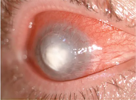
Ayşe Bozkurt Oflaz, Banu Bozkurt, Ümit Kamış, Bengü Ekinci Köktekir Corneal Collagen Crosslinking Treatment in a Case with Pneumococcal Keratitis 2017 Jun 1 Turk J Ophthalmol. 2017 Jun 1; 47(3):161-164 Figure 2. PMCID: PMC5468530. License: CC BY.
A clinical photograph showing a severe case of infectious keratitis after corneal cross-linking, with a white infiltrate and diffuse opacity in the central cornea and marked conjunctival injection.
Corneal infiltrate: White nodular infiltrate in the stroma, commonly in the central to paracentral area. May be accompanied by satellite lesions.
Hypopyon: In severe cases, pus accumulation in the anterior chamber.
Corneal edema: Diffuse conjunctival injection and corneal edema.
Epithelial defect: Often accompanied by epithelial defects due to the ongoing epithelial regeneration process after the epi-off procedure.
Corneal perforation: In severe cases, may progress from descemetocele to corneal perforation. Rana et al. reported two cases of corneal perforation.
The prognosis of reported cases varies. In mild cases (e.g., Staphylococcus epidermidis), corrected visual acuity of 20/22 has been achieved after treatment, while in severe cases (e.g., Pseudomonas aeruginosa, MRSA, Acanthamoeba), corneal transplantation was required and vision was significantly limited.
Epi-off procedure: Loss of the epithelial barrier increases the risk of infection2).
Protective contact lens: The bandage CL used postoperatively can become a reservoir for infection.
Fluoroquinolone resistance: Most post-CXL infections have been reported to be fluoroquinolone-resistant.
In eyes with a history of herpes simplex virus, there is a risk of viral reactivation due to UV irradiation, and CXL is contraindicated1). Kymionis et al. reported a case of herpes simplex keratitis after CXL, with geographic epithelial defect, stromal edema, and anterior chamber inflammation appearing on postoperative day 5, and herpes simplex virus DNA detected by PCR.
QDoes transepithelial CXL have a lower risk of infection?
A
Compared to the epithelium-off method, the transepithelial method preserves the corneal epithelium, so theoretically the infection risk is lower. However, Rana et al. reported cases of bacterial keratitis after transepithelial CXL. The epithelial barrier is an important protective factor but does not guarantee complete prevention of infection.
Oral antibiotics: In severe cases, ciprofloxacin 750 mg etc. may be added
Management of corneal perforation: Temporary closure with cyanoacrylate adhesive, conjunctival flap surgery
Penetrating keratoplasty: Therapeutic corneal transplantation may be needed in severe cases. In the series by Farrokhpour et al., 5 out of 6 cases required PK
PACK-CXL is being studied as an adjunctive therapy for infectious keratitis refractory to medical treatment1).
Infection type
Efficacy
Notes
Bacterial (superficial)
High
Most effective1)
Fungal
Moderate
Less effective than for bacteria1)
Acanthamoeba
Limited
Used adjunctively1)
In the initial report by Iseli et al. (2008), progression of corneal melting was halted in all 5 eyes with antimicrobial-resistant infectious keratitis, and emergency corneal transplantation was avoided1). Makdoumi et al. reported that PACK-CXL monotherapy without antibiotics cured bacterial keratitis in 14 of 16 eyes1).
PACK-CXL has been considered for microbial keratitis resistant to standard antimicrobial therapy. Previous reports indicate that it is more effective in superficial bacterial keratitis, while its effect is limited in cases with deep infection or endothelial plaques1).
In cases with deep keratitis or endothelial plaques, the effect of PACK-CXL is limited1). Because UVA energy is absorbed in the anterior corneal stroma, it is difficult to reach deep infections1).
In CXL, photoactivated riboflavin enters an excited state and reacts with environmental oxygen to produce reactive oxygen species (ROS)1). ROS exert bactericidal effects through the following mechanisms:
Inhibition of replication by damage to microbial DNA
Leakage of cellular contents due to damage to the cytoplasmic membrane
Inactivation of enzymes and membrane transport systems1)
Additionally, the cross-linking reinforcement of collagen fibers by CXL makes the corneal stromamore resistant to enzymatic degradation, suppressing the progression of microbial collagen melting1).
Despite the antimicrobial effect of CXL, infection may occur due to the following reasons:
Loss of epithelial barrier: In the epi-off method, the corneal epithelium is completely removed, resulting in the loss of the most important physical barrier against bacterial invasion.
Reduced tear defense: Antibacterial factors in tears such as phospholipase A2 are diminished with long-term contact lens use.
Postoperative immunosuppression: Local immunosuppression due to steroid eye drops.
Delayed epithelial healing: After CXL, epithelial regeneration may be delayed, prolonging the unprotected period2).
PACK-CXL is attracting attention as a promising adjunctive therapy for infectious keratitis1). While relatively good results have been reported for bacterial keratitis, the effects are inconsistent for fungal infections and deep infections1). To overcome the limitations of efficacy in deep infections, optimization of irradiation protocols is underway.
Regarding the prevention of post-CXL infection, the issue of fluoroquinolone-resistant bacteria is important. It has been pointed out that options other than fluoroquinolones should be considered as postoperative antimicrobial agents. With the spread of transepithelial methods, procedures that preserve the epithelial barrier are increasing, but long-term data are needed on the effect of reducing infection risk.
Lim L, Lim EWL. A review of corneal collagen cross-linking: current trends in practice applications. Open Ophthalmol J. 2018;12:181-213. doi:10.2174/1874364101812010181.
Jhanji V, Ahmad S, Amescua G, et al. Corneal Ectasia Preferred Practice Pattern. Ophthalmology. 2024 Apr;131(4):P205-P246. doi:10.1016/j.ophtha.2023.12.038. PMID:38349299.
Copy the article text and paste it into your preferred AI assistant.
Article copied to clipboard
Open an AI assistant below and paste the copied text into the chat box.