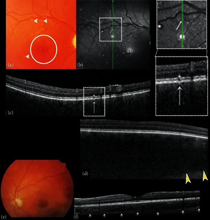
Composite of fundus color photographs and spectral-domain optical coherence (SDOCT) of the right and left eye. (a) The right fundus image shows excavation and pallor of the optic disk, absence of tessellation, diffuse choroidal hemangioma, hypo-hyper pigmentation of the foveal area with absent foveal reflex (circle), and small white dot shaped “microdrusen-like” alterations (arrows). (b) Near-infrared reflectance (NIR) of the right eye shows multiple hyperreflective dots surrounded by a hypo-reflective ring corresponding to the small white dot shaped “microdrusen-like” alterations of the posterior pole observed with ophthalmoscopy corresponded. B-scan cross-sectional SDOCT scan (c) on the hyperreflective dots shows focal alterations of the retinal pigment epithelial (RPE)-photoreceptor lay
Clara Hjalmarsson, Charlotte Backelin, Anders Thoren, Niklas Bergh, Jennifer L. Sloan, Irini Manoli, Charles P. Venditti, Göran Dellgren. Severe heart failure in a unique case of cobalamin-C-deficiency resolved with LVAD implantation and subsequent heart transplantation. Molecular Genetics and Metabolism Reports. 2024;39:101089. doi:10.1016/j.ymgmr.2024.101089.
Akar HT, Yıldız H, Öztürk Z, Karakaya D, Sezer A, Olgaç A. Case presentation: a severe case of cobalamin c deficiency presenting with nephrotic syndrome, malignant hypertension and hemolytic anemia. BMC nephrology. 2024;25(1):217. doi:10.1186/s12882-024-03656-1. PMID:38977946; PMCID:PMC11232354.
Ailliet S, Vandenberghe R, Schiemsky T, et al. A case of vitamin B12 deficiency neurological syndrome in a young adult due to late-onset cobalamin C (CblC) deficiency: a diagnostic challenge. Biochem Med (Zagreb). 2022;32(2):020802. doi:10.11613/bm.2022.020802.
Christopher Goyne, Leena Kansal. Pearls & Oy-sters: Late-Onset Cobalamin C Deficiency Presenting With Subacute Combined Degeneration. Neurology. 2023;100(10):486-489. doi:10.1212/wnl.0000000000201695.