VR perimetry (Virtual Reality Perimetry; VRP) is a technique that uses a VR headset to perform visual field testing in an immersive environment.
Visual field testing is central to glaucoma diagnosis. Standard Automated Perimetry (SAP) is the clinical standard, but it requires monocular occlusion, strict head positioning, and maintaining central fixation 1). The EGS 6th Edition (European Glaucoma Society Guidelines) strongly recommends visual field testing for initial evaluation 3). The AAO PPP (American Academy of Ophthalmology Preferred Practice Pattern) also recommends visual field assessment using SAP4).
Glaucoma affects approximately 80 million people worldwide, and this number is expected to increase to 112 million by 2040 2). To address the growing patient population, more convenient visual field testing methods are needed.
The initial prototype of VRP was the PeriScreener developed by Aravind Eye Hospital. It was a low-cost device combining Google Cardboard, two Android devices, and a Bluetooth clicker. As of 2025, more than 10 types of VRP devices exist, including Oculus Quest, TPP, VirtualEye, AVA, VisuALL, Virtual Field, and Radius 1).
QHow is VR perimetry different from conventional visual field testing?
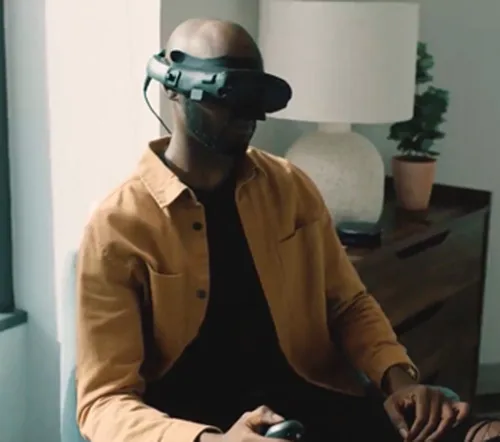
VRP itself is a testing method and differs from subjective symptoms of the disease. The following describes the experiential characteristics of patients undergoing the test.
Immersion: Wearing the VR headset blocks out the surrounding environment.
Operation method: Most devices use a wireless clicker for button operation.
Test duration: Varies by device, but may be equivalent to or shorter than SAP. Virtual Field reports that VRP is on average 76 seconds faster than SAP1).
Claustrophobia: Some patients (with claustrophobia) may experience discomfort2).
Virtual Field (Oculus Go): FDA approved. Background luminance 0.218 cd/m². In 95 eyes, MD r=0.87, PSD r=0.94. Fixation loss rate significantly lower: VRP 0.05 vs SAP 0.13 (p=0.0006), and test time was 76 seconds shorter with VRP1).
Eye tracking / innovative input type
VirtualEye: OLED microdisplay equipped, built-in eye tracking. Implements Visual Grasp mode (detects changes in gaze direction and records responses without button operation)1).
EEG/BCI-based type
nGoggle: Equipped with a Brain Computer Interface (BCI) using electroencephalography (EEG). It detects visual field responses via multifocal steady-state visual evoked potentials (mfSSVEP), eliminating patient-side operational errors.
QWhich VR perimetry device is the most widely used?
A
As of 2025, Virtual Field, which has received FDA approval, and VisuALL, which allows simultaneous binocular testing, are attracting attention. However, due to a lack of standardization among devices, the device adopted varies by facility1).
3. Challenges of Conventional Perimetry and Background of VRP
Detection sensitivity for early glaucoma: Tends to be less sensitive than SAP for mild lesions2).
Limited luminance range: Many devices have a display luminance maximum that differs from the HFA background luminance (31.5 asb ≈ 10 cd/m²)2).
Insufficient eye tracking: Some devices do not adequately detect fixation loss2).
Lack of standardized protocols: Cannot be compared between devices1, 2).
Unfamiliarity with technology: Elderly patients not accustomed to VR technology may have difficulty operating it2).
QCan early glaucoma be missed with VR perimetry?
A
In mild glaucoma, accuracy tends to decrease. Although severity classification shows high agreement (κ = 0.91–0.93), further validation is needed for early detection 1). When early glaucoma is suspected, combined use with SAP is recommended.
In a meta-analysis of 14 studies, the MD correlation between VRP and SAP was generally good, ranging from r=0.77 to 0.941). Only about half of the studies performed Bland-Altman analysis1).
VRP tends to underestimate MS and defect size in glaucoma and overestimate them in healthy individuals1). It has also been reported that accuracy decreases with higher severity1).
Binocular testing with VisuALL has the advantage that the eye with poor fixation can be assisted by fixation of the healthy eye 5).
Slagle et al. (2025) reported a patient with congenital glaucoma (age 23) who had a central scotoma in the left eye 5). Although HFA only yielded unreliable results over 5 years, binocular testing with VisuALL successfully obtained reproducible visual fields of the left eye.
MD (Mean Deviation): Average sensitivity deviation of the entire visual field. Larger negative values indicate more severe sensitivity loss.
PSD (Pattern Standard Deviation): An index reflecting localized sensitivity loss.
VFI (Visual Field Index): Weighted visual field remaining rate centered on the central area (expressed as %).
GHT (Glaucoma Hemifield Test): An index evaluating symmetry between the upper and lower hemifields.
QCan VR perimetry results be compared with conventional test results?
A
MD correlation is good (r=0.77–0.94), but standardization between devices is insufficient 1). Follow-up with the same device is desirable; if changing devices, consider re-establishing baseline.
Conventional SAP presents static stimuli at fixed positions within a bowl perimeter (Goldmann type) and measures threshold sensitivity by varying luminance.
VRP reproduces similar stimulus presentation within a head-mounted display. Background luminance varies widely by device (0.05–25 cd/m²), and many devices differ from SAP’s HFA (31.5 asb ≈ 10 cd/m²) 1). Most devices use the Goldmann III stimulus 1).
The following threshold strategies are used 1).
Full threshold (4/2 dB staircase): Same algorithm as conventional SAP.
ZEST (Zippy Estimation by Sequential Testing): Fast threshold estimation using Bayesian inference.
RATA-Standard: A separately developed estimation strategy.
In VisuALL, each eye has an independent display, and targets are presented alternately. The Dynamic Matrix algorithm compensates for mild convergence insufficiency5). Its application for detecting non-organic visual impairment has also been proposed 5).
The background luminance of Sb-C (0.05 cd/m²) is very low, which may prevent proper measurement of the hill of vision in the macula1). Caution is needed because the luminance characteristics of each device affect interpretation of results.
A 2025 systematic review (14 studies, 10 devices) concluded that VRP shows strong potential for glaucoma visual field assessment 1). However, the lack of test-retest reproducibility data is identified as the major challenge 1).
Hekmatjah et al. (2025) systematically reviewed 14 studies and confirmed generally good agreement between VRP and SAP (MD correlation r=0.77–0.94) 1). Only about half of the studies performed Bland-Altman analysis, and standardization across devices was noted as insufficient.
The EGS 6th Edition mentions the potential of home monitoring using mobile apps 3). The concept of utilizing frequent home VRP test data for trend analysis has also been proposed 6). The Japan Glaucoma Society Guidelines for Glaucoma (5th Edition) also emphasize the importance of visual field testing 6).
The following are listed as research needed in the future 2).
Longitudinal non-inferiority trials: Accumulation of long-term comparative data with SAP.
Technical improvements: Improvement of eye-tracking accuracy and expansion of luminance range.
Development of standardized protocols: Establishment of unified standards enabling cross-device comparisons.
Publication of normative databases: Accumulation of data from diverse races and age groups.
QWill VR perimetry become a standard test in the future?
A
Technical improvements, standardization of protocols, and accumulation of longitudinal studies are necessary1, 2). It holds potential for home monitoring and telemedicine, and the EGS 6th Edition also recognizes this possibility3). At present, it has not completely replaced SAP, but it is expected to become more widespread as a complementary test.
Hekmatjah N, Chibututu C, Han Y, Keenan JD, Oatts JT. Virtual reality perimetry compared to standard automated perimetry in adults with glaucoma: A systematic review. PLoS One. 2025;20(1):e0318074. doi:10.1371/journal.pone.0318074.
Babel AT, Soumakieh MM, Chen AY, Wong C, R da Costa D, Almeida DRP. Virtual Reality Visual Field Testing in Glaucoma: Benefits and Drawbacks. Clinical ophthalmology (Auckland, N.Z.). 2025;19:933-937. doi:10.2147/OPTH.S511803. PMID:40125480; PMCID:PMC11926061.
Pazos M, Traverso CE, Viswanathan A; European Glaucoma Society. European Glaucoma Society - Terminology and guidelines for glaucoma, 6th Edition. Br J Ophthalmol. 2025;109(Suppl 1):1-212. doi:10.1136/bjophthalmol-2025-egsguidelines. PMID:41026937.
Gedde SJ, Vinod K, Wright MM, et al. Primary Open-Angle Glaucoma Preferred Practice Pattern. Ophthalmology. 2021 Jan;128(1):P71-P150. doi:10.1016/j.ophtha.2020.10.022. PMID:34933745.
Slagle GT, Groth SL, Donahue SP, Sponsel WE. Virtual reality perimetry facilitates visual field evaluation in a previously non-assessable eye with severe glaucoma. Am J Ophthalmol Case Reports. 2025;40:102430.