Optical biometry is a test that uses optical interference to measure axial length, corneal curvature, anterior chamber depth, lens thickness, and other biometric data of the eye non-invasively.
Most devices are equipped with swept-source optical coherence tomography (SS-OCT), enabling highly accurate and reproducible measurements. Its main applications include not only IOL power calculation but also obtaining axial length correction values for glaucomaOCT analysis in highly myopic eyes, preoperative refractive surgery evaluation, and follow-up of myopia control treatment with low-concentration atropine.
When Harold Ridley performed the first IOL implantation in 1949, the patient had a refractive error of about 20 D. In the late 1960s, IOL power estimation using vergence formulas began, marking the starting point of modern calculation methods 1). In the 1970s, ultrasound A-mode biometry was established, and later the IOLMaster using partial coherence interferometry (PCI) appeared, standardizing optical measurements. In recent years, third-generation devices with SS-OCT have become widespread, achieving further improvements in accuracy.
QWhat does biometry measure?
A
It measures axial length (AL), corneal refractive power (K value), anterior chamber depth (ACD), lens thickness (LT), and corneal diameter (white-to-white: WTW). These parameters are used to predict the effective lens position (ELP) and calculate the required IOL power. Some devices can also measure central corneal thickness (CCT).
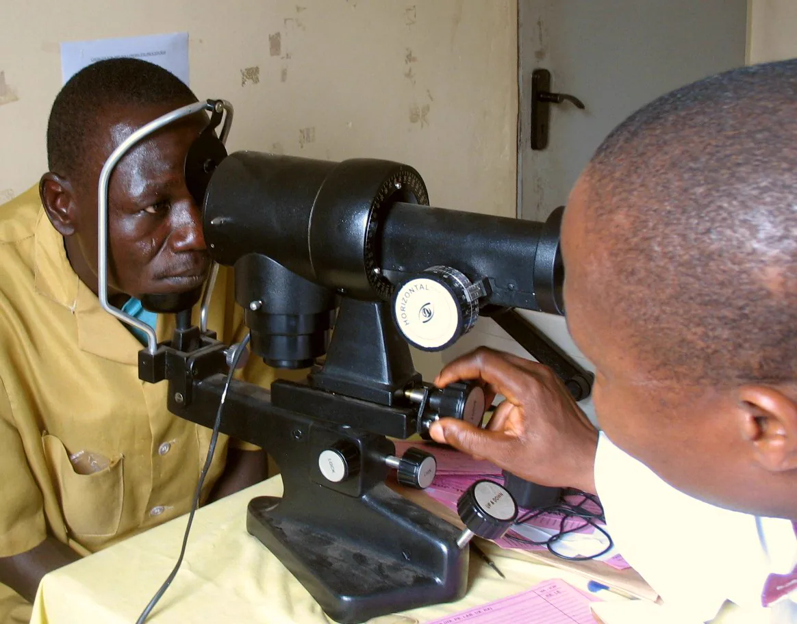
Blyth M. Eye specialist Dr. Ahmedu examining a patient with a keratometer. Figure 1. Source ID: Wikimedia Commons / Eye_doctor_examining_Nigerian_patient_with_keratometer.jpg. 2007. License: CC BY-SA 3.0.
An ophthalmologist measures a patient’s corneal curvature with a keratometer to determine the IOL power needed after cataract surgery. This corresponds to the measurement of corneal curvature (K value) discussed in section “2. Measurement Parameters and Devices.”
Features: Integrates with the cataract surgery support system “CALLISTO eye.” Provides centering guidance for toric IOLs and multifocal IOLs.
Strengths: Adaptability to advanced cataracts and posterior uveitis cases. With swept-source technology, it can measure more cataract eyes than the previous generation PCI 3).
Features: Integration with the astigmatic axis alignment system “VERION.” Implements segmented axial length measurement (applying individual refractive indices to each segment).
Strengths: Segmented correction in long and short eyes is expected to improve calculation accuracy. VERION integration enhances intraoperative astigmatic axis management.
OLCR (1st generation): IOLMaster 500 using partial coherence interferometry (PCI). Measures axial length, K values, and anterior chamber depth.
SS-OCT type (2nd to 3rd generation): Represented by IOLMaster 700 and ARGOS. Uses swept-source laser with wavelength 1,050–1,310 nm. High penetration depth, capable of handling advanced cataracts that were difficult with previous generations.
Optical biometry has been shown to provide significantly higher accuracy and operator-independent results compared to A-mode ultrasound3).
Optical AL error: 0.01–0.02 mm
Ultrasound AL error: 0.1–0.2 mm
When using IOLMaster, adopt measurements with signal-to-noise ratio (SNR) ≥ 5. When using optical biometers, use optical-specific IOL constants. The A-constants provided by IOL manufacturers are only recommended values; optimization based on surgeon’s experience or use of the ULIB database (User Group for Laser Interference Biometry) is beneficial3). Measuring and comparing axial lengths of both eyes helps in early detection of measurement errors.
Cases where the macula is located on the sloping wall of a posterior staphyloma (beware of double peaks)
For these cases, A-mode ultrasound biometry is used. The ESCRS guidelines recommend using ultrasound biometry when optical methods are not applicable for mature or advanced cataracts1).
QWhat to do when optical biometry cannot measure?
A
In cases of dense cataract or poor fixation, optical measurement may be difficult. The alternative is A-mode ultrasound biometry, with the immersion method recommended over the applanation method due to less compression error. Some reports indicate no statistically significant difference between immersion A-scan performed by an experienced operator and optical biometry1).
In ultrasound methods, the sound velocity in the medium directly affects measurement accuracy.
Lens and cornea: approximately 1,641 m/s
Aqueous humor and vitreous body: 1,532 m/s
Average for normal phakic eyes: 1,555 m/s
Contact (applanation) methods compress the cornea, artificially shortening the axial length. Immersion methods avoid compression errors because the probe does not touch the cornea directly, but require careful alignment. Reports indicate that immersion methods performed by experienced operators show no statistically significant difference from optical methods 1).
IOL power calculation formulas have evolved over generations, and currently new-generation formulas such as Barrett Universal II, Kane, and Hill-RBF demonstrate high predictive accuracy. The main characteristics of each formula are as follows 3).
Formula Category
Representative Formula
Additional Variables
Indications
Third Generation (Old Generation)
SRK/T, Holladay I, Hoffer Q
None/ACD
Normal eyes (new generation recommended currently)
Fourth generation
Barrett Universal II, Haigis
ACD, LT, WTW
Good across all axial lengths
AI/Regression hybrid
Kane, Hill-RBF, Pearl-DGS
ACD, LT, WTW
Improved accuracy especially in extreme axial lengths
Older generation regression formulas (SRK-II, SRK, Binkhorst, etc.) are no longer recommended 5). Newer formulas (such as Barrett Universal II) have been reported to improve accuracy, especially in eyes with extreme axial lengths 4).
Post-refractive surgery eyes: Because the anterior-to-posterior corneal curvature ratio is altered, standard formulas produce systematic errors. Specialized methods such as the ASCRS online calculator, Barrett True-K formula, and Haigis-L formula are required 1).
Toric IOLs: Indications include corneal astigmatism of ≥2.0 D with-the-rule or ≥1.5 D against-the-rule. Use of the Haigis-T, Barrett Toric, or Kane Toric formula is recommended 3).
Silicone oil-filled eyes: Optical biometry is most accurate. Because silicone oil acts as a minus lens, the IOL power should be adjusted by 3–5 D 1).
QWhy is IOL calculation difficult in cataract surgery for eyes that have undergone refractive surgery?
A
Refractive surgery (LASIK, PRK, RK) alters the anterior-to-posterior corneal curvature ratio. Keratometers estimate the posterior curvature from the anterior curvature alone, so they overestimate corneal power in post-refractive eyes. Additionally, many IOL formulas predict ELP from axial length and corneal power, but this relationship is altered after refractive surgery, leading to errors. Use of specialized methods (e.g., ASCRS online calculator) is recommended 1).
SS-OCT (Swept-Source Optical Coherence Tomography) measures each ocular interface with high precision based on the following principle.
Light source: Uses a swept-source laser with a wavelength of 1,050–1,310 nm
Interference: Reflected light from each ocular interface (anterior and posterior cornea, anterior and posterior lens, retina) interferes with a reference beam
Calculation: Fourier transform calculates the depth of each interface with high precision
Output: Parameters such as axial length, ACD, LT, and AL are obtained simultaneously
This is the first-generation optical measurement principle adopted by the IOLMaster 500. It measures axial length using the interference pattern of a double beam. Compared to SS-OCT, it has lower penetration and is more likely to be unable to measure in eyes with advanced cataracts.
Conventional optical methods apply a uniform refractive index to the entire eye, which tends to cause overestimation in highly myopic eyes (AL ≥ 25 mm). The “segmented axial length measurement” implemented in ARGOS applies individual refractive indices to each segment (aqueous humor, lens, vitreous body). In long eyes, it displays approximately 0.50 mm smaller than conventional methods, and many calculation formulas have reported improvements in MAE (mean absolute error). However, evidence for this method is still accumulating, and future clinical studies are awaited.
AI-based formulas such as the Hill-RBF method (artificial intelligence pattern recognition), Kane formula, and Pearl-DGS formula have shown improved accuracy 4). Suzuki et al. (2025) conducted a retrospective evaluation of 80 eyes with extreme axial myopia (axial length ≥30.0 mm) and reported that the Kane and Hill-RBF formulas had significantly lower mean absolute errors (MAE) compared to the conventional SRK/T formula 7).
The percentage of eyes within ±0.5 D was 26.3% for SRK/T, 45.0% for Barrett Universal II, 55.0% for Hill-RBF, and 65.0% for Kane. In the subgroup with axial length ≥32 mm, Hill-RBF MAE was 0.49 D and Kane MAE was 0.44 D, which were the best results 7).
Ray tracing based on anterior segment OCT data (Anterion-OKULIX) has been reported to have a significantly lower arithmetic prediction error (−0.13 D vs −0.32 D) compared to the Barrett True K no-history formula in eyes after myopic LVC 6). By directly using full corneal shape data, it is expected to have a theoretical advantage in application to eyes after refractive surgery.
Intraoperative wavefront aberrometry using devices such as the Optiwave Refractive Analyzer is attracting attention as a means to complement preoperative biometry. Reports indicate that in routine adult cataract surgery, it achieves postoperative outcomes comparable to conventional biometry and may enable intraoperative correction of refractive errors 2).
Regular axial length measurements using optical biometers are applied to evaluate the efficacy of myopia control treatments such as low-dose atropine eye drops and orthokeratology. Monitoring axial length every 6 months to 1 year allows objective assessment of treatment effects. Specific measurement intervals and thresholds await future guideline development.
ESCRS Clinical Guidelines. Cataract Surgery Guidelines. European Society of Cataract and Refractive Surgeons; 2023.
Rathod A, Khokhar S, Rani D. Pediatric IOL power calculation: Factors and considerations. Indian J Ophthalmol. 2025;73(3):312-319. doi:10.4103/ijo.ijo_1205_24.
Miller KM, Oetting TA, Tweeten JP, Carter K, Lee BS, Lin S, et al. Cataract in the Adult Eye Preferred Practice Pattern. Ophthalmology. 2022;129(1):P1-P126. doi:10.1016/j.ophtha.2021.10.006. PMID:34780842.
Abbondanza M, Stifani G, Abbondanza D, Leuzzi M. Artificial intelligence applications and cataract surgery. J Clin Med. 2022;11:3899.
Wang L, Koch DD. Intraocular lens power calculations in eyes with previous corneal refractive surgery: review. In: ESCRS Guidelines on Prevention, Investigation, and Management of Post-operative Endophthalmitis and Cataract Surgery. 2024. (ESCRS Cataract Guideline, Section 6.3)
Suzuki Y, Kamoi K, Uramoto K, Ohno-Matsui K. Artificial intelligence driven intraocular lens power calculation in extreme axial myopia. Scientific reports. 2025;15(1):36921. doi:10.1038/s41598-025-20899-6. PMID:41125680; PMCID:PMC12546796.
Copy the article text and paste it into your preferred AI assistant.
Article copied to clipboard
Open an AI assistant below and paste the copied text into the chat box.