Protocol Tでは1年時点においてアフリベルセプト群の視力改善が最大であった。ただし軽度群(最高矯正視力(BCVA) ≧ 20/40)では3剤間に統計的有意差はなかった(8.0 vs 7.5 vs 8.3文字、P > 0.50)11)。
英国の実臨床研究(DRAKO研究)では以下が示された3)。
初回5回の月次投与を完了した患者では12ヶ月で4.2文字改善(全体平均2.5文字改善)
非医師による投与が57%を占めたが安全性は同等であった
DAM Studyでは、アフリベルセプト+マイクロパルスレーザー(MPL)とアフリベルセプト+シャムレーザーを比較した7)。48週時点の注射回数は両群で同等(8.5回 vs 7.9回)であり、MPL追加による最高矯正視力有意差は認められなかった。中心網膜厚(CMT)変化は433 → 288 μmであった7)。
24週:アフリベルセプト群の58.4%がDRSS 2段階以上の改善 vs コントロール群6.0%11)
52週:2q16群65.2%、2q8/PRN群79.9% vs 15.0%が改善11)
100週:視力を脅かす合併症+CI-糖尿病黄斑浮腫の発生率は2q16群16.3%、2q8/PRN群18.7% vs 50.4%11)
Protocol W試験では2年時点でアフリベルセプト群のCI-糖尿病黄斑浮腫+視力低下または増殖糖尿病網膜症(PDR)発生率が16.3% vs シャム群43.5%(HR = 0.32)であった。4年累積では33.9% vs 56.9%(HR = 0.40)と、長期的な疾患進行抑制効果が示された11)。
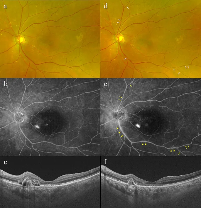
Hidetaka Matsumoto; Junki Hoshino; Saki Numaga; Kaori Mimura; Yosuke Asatori; Hideo Akiyama. Retinal vasculitis after intravitreal aflibercept 8 mg for neovascular age-related macular degeneration. Jpn J Ophthalmol. 2024 Aug 20; 68(5):531-537 Figure 2. PMCID: PMC11420316. License: CC BY.
Images of the left eye of a 79-year-old woman with treatment-naïve neovascular age-related macular degeneration associated with polypoidal choroidal vasculopathy. At baseline, best-corrected visual acuity (BCVA) was 0.8 (0.10 logarithm of the minimum angle of resolution (logMAR) units). (a) Color fundus photograph shows retinal pigment epithelium (RPE) degeneration at the macular area. The retinal vessels appear normal. (b) Fluorescein angiography demonstrates mild leakage and window defects at the macular area. The retinal vessels appear normal. (c) Optical coherence tomography (OCT) shows a shallow irregular RPE elevation (double layer sign) and protruding RPE detachment, reflecting a branching neovascular network and polypoidal lesion, accompanied by subretinal and sub-RPE fluid. The fo
Lopez Fontanet J, Ayala Rodriguez SC, Oliver AL. Aflibercept for the treatment of macular edema secondary to idiopathic retinal vasculitis, aneurysms, and neuroretinitis syndrome. Cureus. 2023;15(4):e38154.
Sivaprasad S, Ghanchi F, Kelly SP, et al. Evaluation of standard-of-care intravitreal aflibercept treatment practices in patients with diabetic macular oedema in the UK: DRAKO study outcomes. Eye (Lond). 2023;37(12):2527-2534.
Neaca R, Manasia D, Tob ME. Uncommon therapeutic approaches for patients with recurrent central serous chorioretinopathy: case report and literature review. Rom J Ophthalmol. 2025;69(1):17-21.
Takahashi N, Kunikata H, Yasuda M, et al. Polypoidal choroidal vasculopathy in a case of retinitis pigmentosa, successfully treated with intravitreal aflibercept. Am J Ophthalmol Case Rep. 2021;23:101123.
Davoudi S, Roohipourmoallai R, Guerin CM, Iyer SSR. Exacerbation of pigment epithelial detachment following aflibercept: a case of bevacizumab rescue. Am J Ophthalmol Case Rep. 2021;24:101216.
Koushan K, Eshtiaghi A, Fung P, Berger AR, Chow DR. Treatment of diabetic macular edema with aflibercept and micropulse laser (DAM Study). Clin Ophthalmol. 2022;16:1109-1115.
Meng PP, Lin CJ, Hsia NY, et al. Use of ultra-widefield fluorescein angiography to guide the treatment to idiopathic retinal vasculitis, aneurysms, and neuroretinitis—case report and literature review. Medicina (Kaunas). 2022;58(10):1467.
Huang YT, Lin CJ, Hsia NY, et al. Intravitreal aflibercept combined with transpupillary thermotherapy in the treatment of refractory macular edema due to primary uveal melanoma. Taiwan J Ophthalmol. 2022;12(4):482-486.
Flaxel CJ, Adelman RA, Bailey ST, et al. Age-Related Macular Degeneration Preferred Practice Pattern. Ophthalmology. 2024.
Flaxel CJ, Adelman RA, Bailey ST, et al. Diabetic Retinopathy Preferred Practice Pattern. Ophthalmology. 2024.
Flaxel CJ, Adelman RA, Bailey ST, et al. Retinal Vein Occlusions Preferred Practice Pattern. Ophthalmology. 2024.