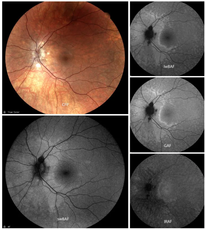
Matthias M Mauschitz; Markus Zeller; Pradeep Sagar; Suchitra Biswal; Gabriela Guzman; Jan H Terheyden. Fundus Autofluorescence in Posterior and Panuveitis—An Under-Estimated Imaging Technique: A Review and Case Series. Biomolecules. 2024 Apr 25; 14(5):515 Figure 5. PMCID: PMC11118036. License: CC BY.
A case of presumed active atypical acute zonal occult outer retinopathy (AZOOR) on color fundus photography (CFP), short-wavelength blue-light autofluorescence (swBAF, 450 nm), long-wavelength blue-light autofluorescence (lwBAF, 488 nm), green-light autofluorescence (GAF, 518 nm), and infrared-light autofluorescence (IRAF, 787 nm). While it is difficult to detect lesions on CFP, they can easily be visualized on different FAF modalities as a hyperautofluorescent pattern expanding from the papilla on swBAF, lwBAF, and GAF, and as a hypoautofluorescent area on IRAF. Source: [13].
Roy R, Dutta Majumder P. Current understanding of acute zonal occult outer retinopathy (AZOOR). Indian J Ophthalmol. 2024;72(7):935-7.
Ünlü BH, Karti O, Saatci AO. A case of an acute zonal occult outer retinopathy variant characterized with an insidious peripheral onset and centripetal progression. Cureus. 2024;16(5):e59600.
Khan S, Saigal K, Moxam J, Maleki A. A case of concomitant acute zonal occult outer retinopathy and secondary nonparaneoplastic autoimmune retinopathy. Case Rep Ophthalmol. 2025;16(1):124-32.
Iuliano M, Lombardo M, Falsini B, Sebastiani J, D’Ambrosio M, Martelli F, et al. Structural, functional, and cellular analysis of a case of acute zonal occult outer retinopathy (AZOOR). Biomedicines. 2025;13(7):1521.
Herbort CP Jr, Arapi I, Papasavvas I, Mantovani A, Jeannin B. Acute zonal occult outer retinopathy (AZOOR) results from a clinicopathological mechanism different from choriocapillaritis diseases: a multimodal imaging analysis. Diagnostics. 2021;11(7):1184.
Karska-Basta I, Romanowska-Dixon B, Pojda-Wilczek D, Bakunowicz-Lazarczyk A, Kubicka-Trzaska A, Gerba-Górecka K. Acute zonal occult outer retinopathy in a patient suffering from epilepsy: five-year follow-up. Medicina. 2021;57(11):1276.
Ahmed Y, Sayal A, Kaplan AJ, Micieli JA. Monocular temporal hemianopia due to acute zonal occult outer retinopathy. Case Rep Ophthalmol. 2022;13(1):44-9.
Özyol E, Özyol P. Acute zonal occult outer retinopathy: optical coherence tomography angiography findings and treatment response. GMS Ophthalmol Cases. 2022;12:Doc16.
Fung AT, Lo-Cao E, Cornish EE. Acute zonal occult outer retinopathy-like presentation secondary to scleral buckle. Am J Ophthalmol Case Rep. 2022;28:101716.
Milovanova E, Fielden MP, Kassam F. Acute zonal occult outer retinopathy in a patient with retinitis pigmentosa. Digit J Ophthalmol. 2022;28.