視神経管は視神経(髄膜に包まれる)と眼動脈を頭蓋窩へ通す管である。上眼窩裂は全長約22mmで蝶形骨大翼と小翼の間に形成され、総腱輪(Annulus of Zinn)により上部・中部・下部に分かれる。上部には涙腺神経(V1)・前頭神経(V1)・滑車神経(IV)・上眼静脈が走行し、中部には鼻毛様体神経(V1)・外転神経(VI)・動眼神経(III)上枝・下枝が走行する。下部には下眼静脈が走行する。この狭い空間に多数の神経・血管が集中するため、眼窩先端部の病変が広範な脳神経障害を引き起こす。
Cavernous sinus thrombosis with bilateral orbital vein involvement and diffuse ischemic retinopathy. Arq Bras Oftalmol. 2023 Jan-Feb; 86(1):79-82. Figure 3. PMCID: PMC11826674. License: CC BY.
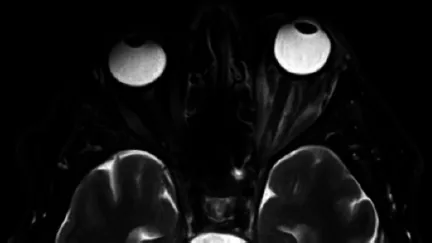
Axial T2-weighted magnetic resonance image of the orbits at 2 weeks, with improvement of the left superior ophthalmic vein thrombus and bilateral cavernous sinus thrombosis.
Mehta S, Gupta K, Patel Nakshiwala N. Orbital apex syndrome due to Aspergillus flavus infection in immunocompetent patients: a report of two cases. Cureus. 2023;15(8):e43508.
Plasencia M, McQueen BR. Orbital apex syndrome secondary to herpes zoster ophthalmicus: a case of irreversible optic nerve damage. Cureus. 2023;15(10):e46522.
Ulatowski H, Bequest A, Sharma A, et al. Granulomatosis with polyangiitis presenting as orbital apex syndrome. Cureus. 2024;16(7):e64087.
Rajad H, Bigi S, Adnor S, et al. Orbital apex syndrome associated with cranial nerve V neuritis complicating bacterial maxillary sinusitis. Radiol Case Rep. 2025;20:3859-3864.
Marzoughi S, Chen T. Orbital apex syndrome due to mucormycosis - missed on initial MRI. Neurohospitalist. 2022;12(1):127-130.
Zielke T, Kim M, Simon JE, et al. Hypertrophic cranial pachymeningitis and orbital apex syndrome secondary to infection of the eye: illustrative case. J Neurosurg Case Lessons. 2021;1(21):CASE20168.
Fukushima A, Mihoshi M, Shimizu Y, et al. A case of orbital apex syndrome related to herpes zoster ophthalmicus. Cureus. 2022;14(7):e27254.
Eldweik L. Radiation induced tissue necrosis mimicking orbital apex syndrome. SAGE Open Med Case Rep. 2022;10:2050313X221123292.
Massey D, Saab M. Orbital apex syndrome secondary to SMARCB1-deficient invasive sinonasal carcinoma. Cureus. 2022;14(11):e31017.